|

|
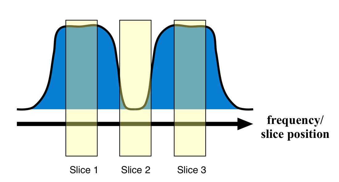
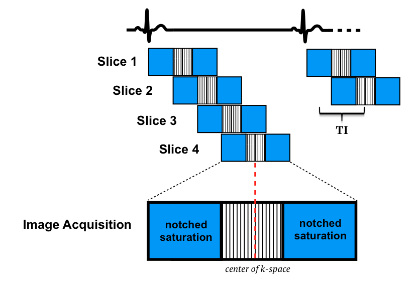
A clever method to permit lengthening of TI with little or no penalty in the number of slices is to use slice-selective notched RF-pulses. Notched RF-pulses saturate adjacent slices but have a mid-frequency gap leaving the center slice unaffected. TI is nearly doubled as the saturation pulse for a given slice has occurred when the previous slice was imaged. At present, this option is available only on GE scanners.
|
Advanced Discussion (show/hide)»
Further Details about RF-pulses
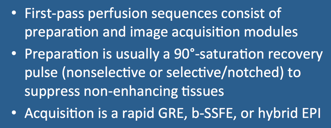
Three broad choices are available for the type of preparation pulses used.
Non-selective RF-pulses are most widely used in first-pass imaging. The term "non-selective" means that these pulses affect the entire volume of interest (e.g., the heart) and are not confined to a single slice. They are also known as "hard" pulses, being recognized by their a rectangular shape. A major advantage of non-selective pulses is their short duration (<3 ms). Crusher gradients are commonly applied after the pulses to minimize off-resonance artifacts and stimulated echoes from T2-coherences.
A second advantage of non-selective pulses is the ability to perform multi-planar imaging. With this saturation method, for example, it is possible in a single acquisition to generate 3 short-axis views and 2 long-axis views simultaneously.
Adiabatic pulses. The 90º-crusher combination is sensitive to B1 field variations, so composite adiabatic RF-pulses (like BIR-4) have been used to create more uniform saturation effects. However, these pulses take longer to perform and deposit more energy in the tissues (higher SAR).Slice-selective pulses, like the notch pulse described above, have some advantages of increased number of slices for a given TI. The major disadvantage is that multiplanar acquisition is not possible as it is with non-selective pulses. Selective pulses will cause cross-talk saturation interference in other imaging slices. So usually only a set of parallel short-axis views are obtained.
Further Details about Image Acquisition Modules
The principal techniques available all require tradeoffs between signal linearity, signal-to-noise, spatial resolution, temporal resolution, and artifacts.
Spoiled-GRE methods are probably the most popular and widely used at present, especially at 3T. Typical imaging parameters might be TR/TE/FA = 2/1/10° at 150 ms per section. Due to the low flip angle, spoiled-GRE sequences have the lowest signal-to-noise The major advantage of this method is reduced motion and blood flow artifacts.
Balanced-SSFP methods (e.g. FIESTA, TrueFISP) produce the highest signal-to-noise of the major techniques. Typical imaging parameters would be similar to the spoiled-GRE technique but with a much higher flip angle (e.g. 50° instead of 10°). Major disadvantages include off-resonance artifacts ("moiré pattern") and frequency shifts induced by the gadolinium bolus. These features currently restrict the use of b-SSFP sequences to 1.5T and below.
Hybrid echo-planar/gradient echo techniques permit the fastest image acquisition times per slice (100-120 ms) and slightly greater spatial coverage especially at higher heart rates. Faster imaging translates to fewer motion-induced artifacts especially near the endocardium. Typical imaging parameters might be TR/TE/FA = 6/1/25° with an echo train length (ETL) = 4-6. The major disadvantages are the presence of susceptibility artifacts and T2-blurring, both of which make these sequences more challenging to implement at 3T where reduction in ETL is usually required.
Coelho-Filho OR, Rickers C, Kwong RY, Jerosch-Herold M. MR myocardial perfusion imaging. Radiology 2013; 266:701-725.
Ding S, Wolff SD, Epstein FH. Improved coverage in dynamic contrast-enhanced cardiac MRI using interleaved gradient-echo EPI. Magn Reson Med 1998; 39:514-519. (original description of hybrid EPI technique).
Fair MJ, Gatehouse PD, DiBella EVR, Firmin DN. A review of 3D first-pass, whole-heart, myocardial perfusion cardiovascular magnetic resonance. J Cardiovasc Magn Reson 2015; 17:68e
Gerber BL, Raman SV, Nayak K, et al. Myocardial first-pass perfusion cardiovascular magnetic resonance: history, theory and current state of the art. J Cardiovasc Magn Reson 2008; 10:18.
Hunold P, Maderwald S, Eggebrecht, et al. Steady-state free precession sequences in myocardial first-pass perfusion MR imaging: comparison with TurboFLASH imaging. Eur Radiol 2004; 14:409-416
Jung B, Honal M, Hennig J, Markl M. k-t-Space accelerated myocardial perfusion. J Magn Reson Imaging 2008; 28:1080-1085.
Kellman P, Arai AE. Imaging sequences for first pass perfusion − a review. J Cardiovasc Magn Reson 2007; 9:525-537.
Shehata ML, Basha T, Hayeri MR, et al. MR myocardial perfusion imaging: insights on techniques, analysis, interpretation, and findings. RadioGraphics 2014; 32:1636-1657.
Slavin GS, Wolff SD, Gupta SN, Foo TKF. First-pass myocardial perfusion MR imaging with interleaved notched saturation: feasibility study. Radiology 2001; 219:258-163. (The notched RF method).
Wang Y, Moin K, Akinboboye O, Reichek N. Myocardial first pass perfusion: steady-state free precession versus spoiled gradient echo and segmented echo planar imaging. Magn Reson Med 2005; 54:1123-1129.


