Note: In this Q&A we adopt the convention that substances "bright" on T1-weighed images have short T1 values. This is true for most (but not all) pulse sequences. For a detailed explanation, click here.
|
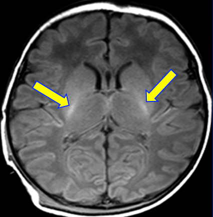
No. But don't feel bad if you thought so. This is an extremly common misconception. The relatively brighter areas of myelinated brain seen on T1-weighted images come entirely from the ¹H nuclei of water, not "fatty myelin" or sphyngolipids.
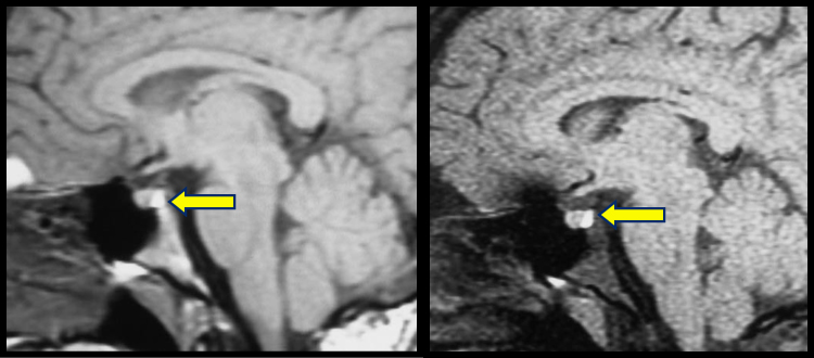
A second example is the commonly seen pituitary "bright spot". In the early days of MRI the pituitary bright spot was misidentified as a sellar "fat pad" because of its high signal on T1 weighted images. I even fell for this trap myself! It was soon recognized the pituitary bright spot was also a water resonance, shortened by association with phospholipid vesicles containing anti-diuretic hormone in the neurohypophysis. For the non-believers, see the pre- and post-fat saturation images below. |
The above are just two common examples of how water molecules closely associated with macromolecules may have their molecular motions slowed so that their T1 values are significantly shortened, so much so that they may be mistaken for fat. Other examples include the very bright neonatal pituitary gland (loaded with endoplasmic reticulum and proteins) and the edges of demyelinating lesions (from water associated with fragmented myelin).
These concepts are explored more completely in several other Q&A's on the website.
These concepts are explored more completely in several other Q&A's on the website.
Advanced Discussion (show/hide)»
No supplementary material yet. Check back soon!
References
Deese AJ, Dratz EA, Hymel L, Fleischer S. Proton NMR T1, T2, and T1ρ relaxation studies of native and reconstituted sarcoplasmic reticulum and phospholipid vesicles. Biophys J 1982; 37:207-216.
Holder CA, Elster AD. Magnetization transfer imaging of the pituitary: further insights into the nature of the posterior “bright spot.” J Comput Assist Tomogr 1997; 21:171-174.
Related Questions
What is T1 relaxation?
Can you explain a little more about the dipole-dipole interaction? I still don't quite understand.
How does the presence of macromolecules affect T1 and T2?
What is T1 relaxation?
Can you explain a little more about the dipole-dipole interaction? I still don't quite understand.
How does the presence of macromolecules affect T1 and T2?


