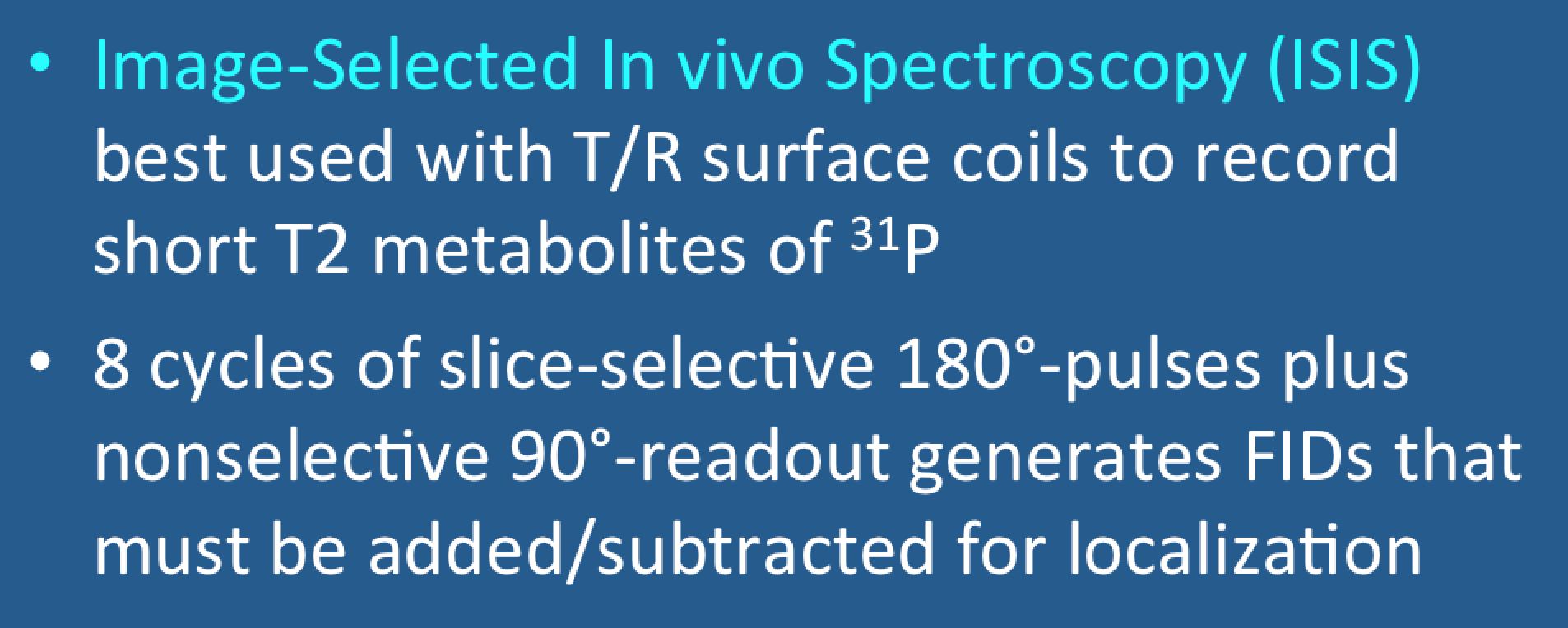
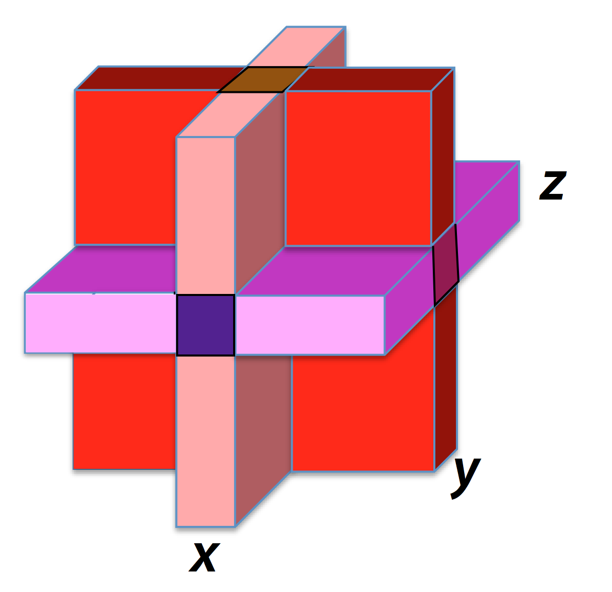
Image-Selected In vivo Spectroscopy (ISIS) was developed in the 1980s and probably remains the best single voxel spectroscopy (SVS) for ³¹P when using transmit/receive surface coils. The recorded signal is a free induction decay (FID). Similar to PRESS and STEAM, ISIS uses a slice intersection strategy for voxel localization. However, the required spectrum is not obtained from a single acquisition, but must be calculated using data from 8 separate RF-pulse cycles. These FIDs are added and subtracted in a particular order to define the volume of interest. A simplified version of the ISIS sequence is shown below:
The sequence includes three slice-selective 180°-inversion pulses and gradients in three orthogonal directions. The 8 separate acquisitions needed to calculate the final ISIS spectrum result from the 2³ = 8 permutations of inversion pulses (off/off/off, off/off/ON, off/ON/ON, etc.). The FID in each cycle is obtained using a nonselective ("hard") 90°-pulse that excites the entire volume under the coil.
To better understand how this works in practice, the diagram below shows how a 2-cycle ISIS sequence in one dimension could be used to localize a single planar slab of data.
To better understand how this works in practice, the diagram below shows how a 2-cycle ISIS sequence in one dimension could be used to localize a single planar slab of data.
The metabolites of interest in ³¹P spectroscopy have relatively short T2 values, and hence ISIS allows capturing more of this signal than either PRESS or STEAM. During the ISIS localization process, the magnetization remains along the longitudinal axis (± z) except during the short readout period of the FID.
The major limitations of ISIS result from subtraction errors. These occur primarily due to signal contamination when various portions of the imaged tissue block respond slightly differently to the RF-pulses. Motion artifacts during the 8-cycle acquisition also contribute.
The major limitations of ISIS result from subtraction errors. These occur primarily due to signal contamination when various portions of the imaged tissue block respond slightly differently to the RF-pulses. Motion artifacts during the 8-cycle acquisition also contribute.
Advanced Discussion (show/hide)»
To improve uniformity of B1 excitation, the RF-pulses used in transmit/receive surface coil implementations of ISIS are typically adiabatic — hyperbolic secant full (180°) or half passage (90°) are good choices. Adiabatic pulses utilize simultaneous amplitude and frequency modulation. They have excellent slice profiles with flat response and no side lobes leading to better edge definition of the voxel.
In addition to 31P spectroscopy, ISIS is also widely used in 15N spectroscopy, whose metabolites, like those of phosphorus, have very short T2 values.
Like PRESS and STEAM, ISIS can also be used in a multi-voxel mode, being combined with 2D or 3D chemical shift imaging techniques with phase-encoding gradients.
References
Ordidge RJ, Connelly A, Lohman JAB. Image selected in vivo spectroscopy (ISIS). A new technique for spatially selective NMR spectroscopy. J Magn Reson 1986; 66:283-294.
Ordidge RJ, Connelly A, Lohman JAB. Image selected in vivo spectroscopy (ISIS). A new technique for spatially selective NMR spectroscopy. J Magn Reson 1986; 66:283-294.
Related Questions
If frequency-encoding cannot be used to determine spatial position, how do you localize an MRS signal?
If frequency-encoding cannot be used to determine spatial position, how do you localize an MRS signal?


