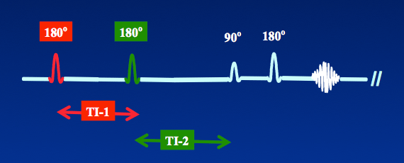
Double Inversion Recovery (DIR) is an inversion recovery variant that uses not one, but two nonselective 180°-inverting pulses. The time associated with each are commonly denoted TI-1 and TI-2. The pulse sequence timing diagram is:
|
To date, most of the applications of this type of DIR technique has been for brain imaging, especially for detection of multiple sclerosis plaques and lesions of the cerebral cortex. Here the first 180°-pulse suppresses CSF and the second suppresses white matter.
|
 Black blood DIR image of heart
A differently structured DIR technique producing "black blood" is commonly used in cardiovascular MRI. In this method, two 180°-pulses are applied very close together in time. The first 180°-pulse is nonselective, meaning that it inverts the magnetization for all slices within the imaging volume. The second 180°-pulse, following immediately on the heels of the first 180°-pulse, is slice selective, meaning it returns the magnetization of all tissues in that slice only back to the +z-direction. Thus the myocardium and other relatively stationary tissues have their signals preserved, but the blood flowing from adjacent slices has an inverted magnetization. The TI is adjusted to null the signal from inflowing blood, which appears "black" on magnitude reconstructed IR images.
Although two 180°-pulses are used, there is really only one material (blood) being inverted and hence only one TI value (measured from the time of the first inverting pulse). Thus this sequence in many ways behaves more like a single IR sequence than the DIR for brain imaging described above.
It is possible to use a third (or even fourth) 180°-pulse in conjunction with a black blood DIR technique. This third pulse is commonly used to suppress pericardial fat, producing a triple inversion recovery (TIR) sequence, that is a combination of STIR plus black blood DIR. These techniques will be described in more detail in a later Q&A.
Advanced Discussion (show/hide)»
No supplementary material yet. Check back soon!
References
Young IR, Hall AS, Bydder GM. Design of a multiple inversion-recovery sequence for T1 measurement. Magn Reson Med 1987; 5:99-108.
Redpath TW, Smith FW. Technical note: use of a double inversion recovery pulse sequence to image selectively grey or white brain matter. Br J Radiol 1994; 67:1258-1263.
Young IR, Hall AS, Bydder GM. Design of a multiple inversion-recovery sequence for T1 measurement. Magn Reson Med 1987; 5:99-108.
Redpath TW, Smith FW. Technical note: use of a double inversion recovery pulse sequence to image selectively grey or white brain matter. Br J Radiol 1994; 67:1258-1263.
Related Questions
Why use IR? What are the advantages?
Isn't double inversion recovery enough? Why would you want to do triple IR?
When is double inversion recovery used for cardiac imaging? How does it work?
Why use IR? What are the advantages?
Isn't double inversion recovery enough? Why would you want to do triple IR?
When is double inversion recovery used for cardiac imaging? How does it work?


