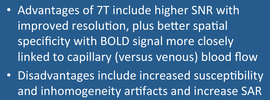
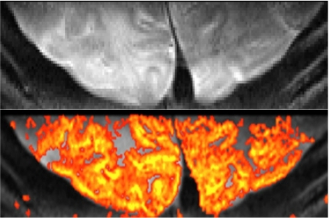
A well recognized benefit of 7T imaging is the concomitant increase in BOLD signal and signal-to-noise ratio (SNR). Improved SNR translates into better spatial resolution, allowing voxels ≤ 1 mm³ to be obtained in reasonable imaging times.
|
Additionally, BOLD signal at 7T more closely reflects the concentration of deoxyhemoglobin in capillaries rather than in larger veins (as it does at 1.5T). This phenomenon results from a field-dependent shortening of blood T2 and T2* relative to brain described in a prior Q&A and in the references below. fMRI at 7T thus offers the potential of improved spatial specificity since cortical neuronal activity is more closely linked to blood flow changes in the microvasculature than in larger vessels. This effect can be further accentuated by using spin-echo (SE) instead of gradient echo (GRE) acquisition.
|
Notwithstanding these advantages, some limitations at 7T are well recognized: 1) increased susceptibility artifacts near the frontal sinuses and skull base (producing spatial distortion and signal drop out); 2) increased dielectric artifacts (manifest by signal inhomogeneity); 3) increased specific absorption rate (SAR, restricting number of slices); and 4) more difficult RF-coil design and shimming (causing inhomogeneous excitation and signal variations).
Advanced Discussion (show/hide)»
The BOLD signal is indeed substantially increased at 7T relative to 3T, but the SNR improvement is somewhat blunted as physiological noise becomes more important than thermal noise at higher fields.
References
Barth M, Poser BA. Advances in high-field BOLD fMRI. Materials 2011; 4:1941-1955.
Harel N. Ultra high resolution at ultra-high field. NeuroImage 2012; 62:1024-1028.
Olman CA, Yacoub E. High-field fMRI for human applications: an overview of spatial resolution and signal specificity. Open Neuroimaging J 2011; 5 (Suppl 1-M3):74-89.
Siero JCW, Ramsey NF, Hooguin H, et al. BOLD specificity and dynamics evaluated in humans at 7T: Comparing gradient-echo and spin-echo hemodynamic responses. PLOS One 2013; 8(1) e354560:1-8.
Uludag, Müller-Bierl B, Ugurbil K. An integrative model for neuronal activity-induced signal changes for gradient and spin echo functional imaging. NeuroImage 2009; 48:150-165.
Related Questions
How is image contrast produced by BOLD fMRI?
How is image contrast produced by BOLD fMRI?

