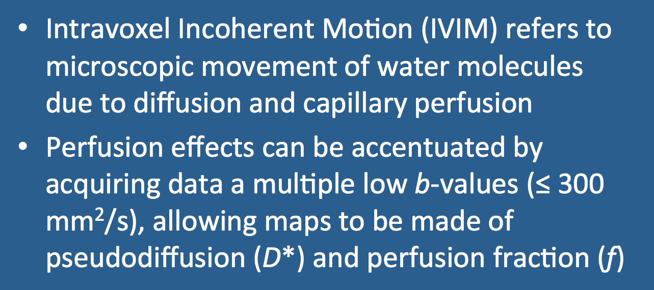
Denis Le Bihan, father of modern diffusion imaging, coined the term intravoxel incoherent motion (IVIM) in the 1980s to refer to the microscopic translation of water molecules within a voxel during an MR experiment. When gradients are applied during evolution of the MR signal, IVIM causes spin dephasing and signal loss.
If the gradients are relatively strong, IVIM-induced signal losses are primarily due to diffusion — the Brownian motion of water molecules in and around cells. When weaker gradients are used, however, a second IVIM mechanism also contributes to signal loss — microcirculation of blood in the capillary network. Microscopic perfusion effects have been largely ignored in the 25 years since Le Bihan's seminal papers, but have recently enjoyed a renaissance in what is now known as IVIM imaging.
If diffusion-only effects are considered, MR signal intensity (S) relative to baseline (So) without diffusion-sensitizing gradients can be expressed as
S = Soe−bD or S/So = e−bD
where D is the apparent (or measured) diffusion coefficient and b is a factor reflecting the strength and duration of the pulsed diffusion gradients. Under this model, a semilogarithmic plot of signal attenuation ln(S/So) vs b should be a straight line with slope = D.
|
When careful signal measurements are made using a large number of different b-values, however, considerable departures from this simplified mono-exponential model are observed. At low b-values (i.e., ≤ 300 − 500 s/mm²) the signal attenuation is greater (and the calculated ADC higher) than expected due to increased IVIM loses from microscopic perfusion. At larger b-values (≥ 1000−1500 s/mm²), signal attenuation is often less than expected, an effect due to the non-Gaussian shape (kurtosis) of the diffusion probability distribution further discussed in the next Q&A.
|
The IVIM model as originally proposed by Le Bihan et al takes into account both diffusion and perfusion effects using the following bi-exponential model
S/So = fe−b(D+D*) + (1−f)e−bD
 Enlarged area of graph showing D* vs D
Here f (dimensionless) is the perfusion fraction, the percent of a voxel volume occupied by capillaries. Conversely (1−f) reflects the extravascular space where only diffusion effects (with apparent diffusion coefficient D) take place. The parameter D* is called the pseudodiffusion coefficient and reflects dephasing due to perfusion in semi-randomly organized capillaries. D* is sometimes referred to as ADCfast or ADChigh while D is sometimes called ADCslow or ADClow. Depending on the steepness of the initial portion of the curve, (which in turn depends on capillary density and perfusion), D* may be 5-10x greater than D.
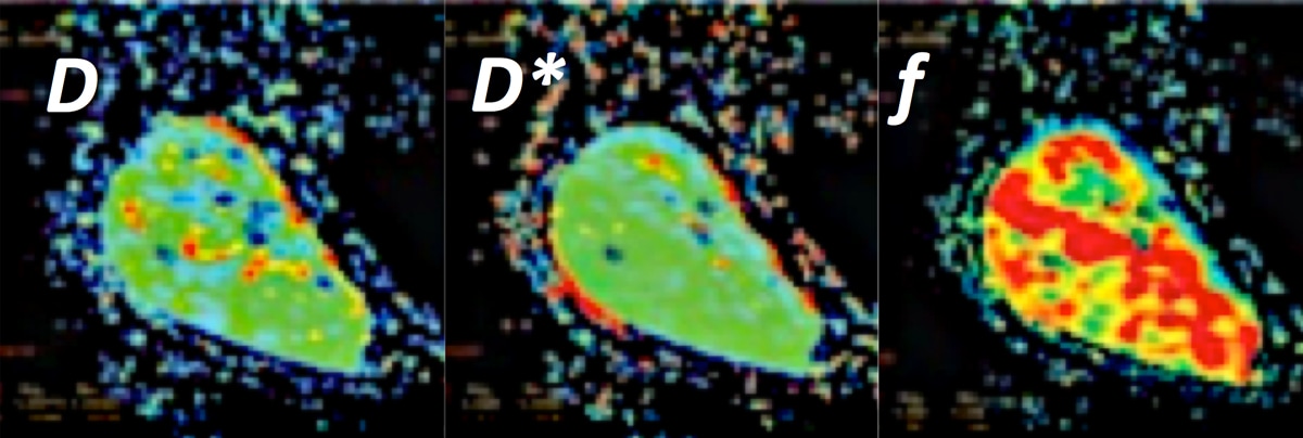
By acquiring DWI images at multiple b-values and fitting the data to above equation, it is possible to estimate f, D, and D* and create maps for each. Typically between 6 and 10 data sets are obtained with b-values ranging between 0 and 1000 s/mm², with at least half the measurements performed at less than 250 s/mm².
Some interesting results of IVIM imaging have been reported for tumors of the prostate, liver, and soft tissues, where perfusion fractions have been shown to correlate with other methods of assessing vascularity. However, considerable errors occur in the estimation of D*, which I personally feel are unreliable, varying too much among centers and studies. Not only D*, but all IVIM parameter estimates are affected by motion, noise, and secretory physiologic processes (such as glandular and renal tubular flows) that cannot easily be distinguished from capillary perfusion. I thus remain a little skeptical for now as to what extent IVIM imaging will find a role in our future imaging armamentarium.
Advanced Discussion (show/hide)»
No supplementary material yet. Check back soon!
References
Du J, Li K, Zhang W, et al. Intravoxel incoherent motion MR imaging: comparison of diffusion and perfusion characteristics for differential diagnosis of soft tissue tumors. Medicine 2015; 94:1-8.
Iima M, Le Bihan D. Clinical intravoxel incoherent motion and diffusion MR imaging: past, present and future. Radiology 2016; 278:13-32.
Koh D-M, Collins DJ, Orton MR. Intravoxel incoherent motion in body diffusion-weighted MRI: reality and challenges. AJR Am J Roentgenol 2011; 196;1351-1361.
Le Bihan D, Breton E, Lallemand D, et al. MR imaging of intravoxel incoherent motions: applications to diffusion and perfusion in neurologic disorders. Radiology 1986; 161:401-407. (This seminal paper describes IVIM, separating perfusion and diffusion effects.)
Le Bihan D, Breton E, Lallemand D, et al. Separation of diffusion and perfusion in intravoxel incoherent motion MR imaging. Radiology 1988; 168:497-505.
Related Questions
What is meant by the b-value? How do I pick it?
What is diffusion kurtosis and how does it differ from regular "diffusion"?
What is meant by the b-value? How do I pick it?
What is diffusion kurtosis and how does it differ from regular "diffusion"?


