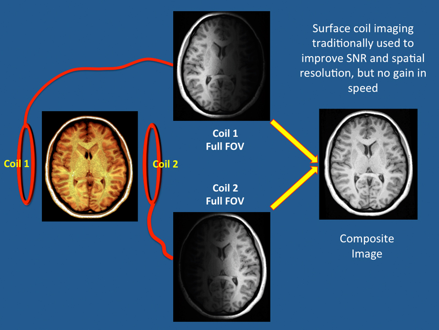
Parallel and "regular" MR imaging share the common feature of multiple receiver coils, but process the information completely differently. Consider the simplified receiver array pictured below consisting of two coils (#1 and #2) placed on either side of the head. As expected, the signal received from each surface coil is strongest nearest the coil and drops off with distance.
|
In "regular" MR imaging we can combine data from each surface coil to produce a composite image of the brain at full field-of-view (FOV). This arrangement may provide improved signal-to-noise and spatial resolution compared to that obtainable from a single large head coil, but produces no gain in speed.
|
|
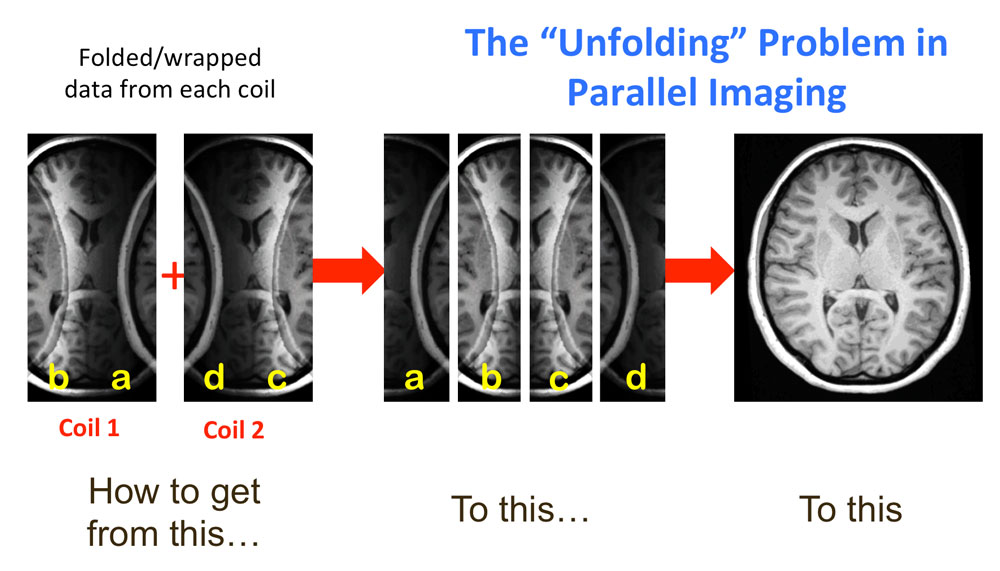
To shorten imaging time in PI the number of phase-encoding steps are reduced. For speed to be doubled at constant resolution, only half of the lines of k-space are acquired. This strategy, however, results in an insufficient number of spatial frequencies collected to adequately represent the imaged object. A reduced FOV and aliasing ("wrap-around") is seen in the reconstructed image.
|
Wrap-around and reduced FOV also occur in data obtained from the individual surface coils (#1 and #2). Routine image reconstruction (as done with "regular" MRI) would result in a mess of overlapping edges and shadows due to uncorrected aliasing. The fundamental problem is PI is how to "unfold" this data using information about the coils themselves.
In the frequency domain, an equivalent problem exists — how to estimate the missing lines of k-space and avoid aliasing wrap-around.
Two basic strategies have been developed to correct for the aliasing overlap in PI images. In the first method (SENSE, ASSET), images from the individual coils are reconstructed and correction is performed in the spatial/image domain. The second group of methods (GRAPPA, ARC) operate in the frequency domain; data is corrected in k-space before reconstruction.
Both techniques are described in detail in subsequent Q&A's.
Both techniques are described in detail in subsequent Q&A's.
Advanced Discussion (show/hide)»
No supplementary material yet. Check back soon!
References
Deshmane A, Gulani V, Griswold MA, Seiberlich N. Parallel MR imaging. J Magn Reson Imaging 2012;36:55-72. (review)
Glockner JF, Hu HH, Stanley DW, et al. Parallel MR imaging: a user's guide. Radiographics 2005;25:1279-1297.
Larkman DJ, Nunes RG. Parallel magnetic resonance imaging. Phys Med Biol 2007;52:R15-R55 [review]
Related Questions
Our scanner has two different options for parallel imaging (SENSE and GRAPPA). What are these and how do they differ?
Our scanner has two different options for parallel imaging (SENSE and GRAPPA). What are these and how do they differ?


