|
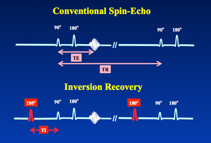
Inversion recovery (IR) is a conventional spin echo (SE) sequence preceded by a 180° inverting pulse. In other words, if a SE sequence is denoted by {90°−180°−echo}, the IR sequence can be written
180° — {90°−180°−echo}
The time between the 180° inverting pulse and the 90°-pulse is called the inversion time (TI). The repetition time (TR) and echo time (TE) are defined as they are for spin echo.
|
 Regrowth of tissue Mz after 180° pulse.
The function of the inverting pulse is to flip the initial longitudinal magnetization (Mo) of all tissues in the imaged slice or volume to point opposite to the direction of the main magnetic field (Bo). During the TI interval, these inverted tissues undergo T1 relaxation as they variably seek to re-establish magnetization along the +z-direction. When spin echo signal generation begins (at the 90°-pulse), the initial longitudinal magnetizations of different tissues are now separated based on their different intrinsic T1 relaxation times. The degree of separation (and hence image contrast) is controlled by varying the TI parameter in the pulse sequence. Additional contrast effects are also obtained by manipulation of TR and TE.
In addition to the timing parameters (TR/TI/TE), how the MR signal is reconstructed has a significant effect on image contrast and appearance. The two reconstruction methods used for IR are magnitude reconstruction and phase-corrected reconstruction, the subject of a separate Q&A.
Historically, IR techniques were widely used in the "early days" of MRI (c. 1980-1985). They produced excellent image contrast, especially for T1-weighted imaging, but were time-consuming (15-20 minute sequences typical). After about 1985 spin-echo techniques largely supplanted IR for most applications. In the late 1990's use of the fast spin echo (FSE) signal generation allowed high quality IR-FSE sequences to be performed in a more reasonable time frame (5-10 min). Today, IR techniques are widely used in all branches of MRI, but especially for neuroradiology and cardiac imaging applications.
Advanced Discussion (show/hide)»
No supplementary material yet. Check back soon!
References
Bydder GM, Young IR. MR imaging: clinical use of the inversion recovery sequence. J Comput Assist Tomogr 1985; 9:659-675.
Bydder GM, Young IR. MR imaging: clinical use of the inversion recovery sequence. J Comput Assist Tomogr 1985; 9:659-675.
Related Questions
What is phase-sensitive IR and why does it have a gray background?
What is phase-sensitive IR and why does it have a gray background?

