In passive shimming small pieces of sheet metal or ferromagnetic pellets are affixed at various locations within the scanner bore to improve homogeneity. Conversely, active shimming uses currents directed through specialized coils to generate a "corrective" magnetic field.
Active shim coils can be: 1) superconducting, located within the liquid helium-containing cryostat; or 2) resistive, mounted on the same support structure as the gradient coils within the room-temperature inner walls of the scanner. Both types of active shims require their own power supplies and are controlled by special circuitry. Some scanners use both types.
Active shim coils can be: 1) superconducting, located within the liquid helium-containing cryostat; or 2) resistive, mounted on the same support structure as the gradient coils within the room-temperature inner walls of the scanner. Both types of active shims require their own power supplies and are controlled by special circuitry. Some scanners use both types.
|
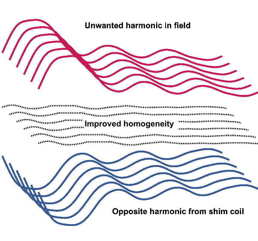
The theory underlying all active shimming methods is based on spherical harmonic analysis, described in a previous question (click here for link). Field mapping first identifies various unwanted harmonic components in the inhomogeneous field. For each unwanted spherical harmonic component in the uncorrected magnetic field, a carefully controlled supplemental magnetic field is generated by passing current through an active shim gradient. This supplemental shim field has the same spatial distribution, but is equal and opposite to the unwanted component. By super-positioning and merging these two opposite magnetic fields together, a neutralization and cancellation of the magnetic field error (inhomogeneity) is effected.
|
Superconducting shim coils are commonly encountered in magnets with fields of 3T or higher. Except for GE Healthcare, however, few manufacturers have used them in lower field strength scanners. Where present, superconducting shim coils (5-20 in number) are embedded in the cryostat just beyond the main coil windings and may correct for several orders of inhomogeneity. Each coil can be individually powered during the shimming process and has a switch allowing it to be placed in persistent superconducting mode once the desired field correction has been obtained. Unlike resistive shims, the current in superconducting shims and the magnetic fields they generate cannot be easily changed once set.

Resistive shim coil tube from a 7T scanner.
Resistive shimming relies on the passage of current through coils located near the room-temperature inner bore of the scanner. For first-order (linear) shimming, additional coils are not needed as the standard x-, y- and z-gradient coils used for imaging can double as shim coils. In a process known as gradient offset shimming the imaging gradients carry a small current (called the offset bias), calculated to reduce residual linear inhomogeneities in the main magnetic field. This method is used in virtually all scanners. It saves space in the patient bore by removing the need for a separate set of first order shim coils.
Some scanners also have additional resistive coils for higher-order shimming. Structurally, these are a series of individual wire windings or conductive patterns etched into copper sheets and formed onto a cylindrical surface. Usually they occupy the space between the primary and secondary gradient coils used for imaging and may be manufactured with the gradients as a single unit. A minimum of 5 separate coils are usually employed to obtain second order shimming; even more are needed for higher orders of field correction.
In the classical shim coil arrangement, one coil is designed to correct each spherical harmonic. Axial coils (typically ring-shaped) correct harmonics in the z-direction (Z, Z2, Z3, etc). Transverse coils (often saddle shaped) correct more complex harmonics (XY, YZ, X2-Y2, ZXY, etc). This arrangement is not optimal in that neighboring windings often carry oppositely running currents. Newer matrix shim coil designs take this into account and are more efficient in reducing the number of windings required.
The big advantage of resistive shims over passive and superconducting ones is that the currents through resistive shims can be changed dynamically. This allows shimming to be performed on a patient-by-patient basis. During the preparatory phase before routine MR scanning begins, rapid automated shimming is now performed routinely on many scanners. More detailed shimming using both automated and manual techniques is required when performing spectral fat suppression and MR spectroscopy.
Advanced Discussion (show/hide)»
The article by Peter Jezzard in the references below gives insight into a variety of additional shimming techniques beyond the standard ones described above. These include: dynamic shimming, local shimming, and acquisition-based methods.
Of these additional techniques, local shimming is most commonly used, especially for the imaging of infants, small parts (hands and feet), and structures that change shape quickly (face and neck). The usual method is to pack saline bags around the object of interest, improving the geometry and reducing susceptibility distortions before shimming is performed. This simple technique increases the uniformity of RF stimulation and is especially helpful for improving spectral fat suppression when employed.
References
Flasche M, Fischer D. White Paper: Magnet homogeneity and shimming. Siemens Healthcare GmbH, 2017.
Gruetter R. Automatic, localised in vivo adjustment of all first- and second-order shim coils. Magn Reson Med 1993; 29:804-811. (This paper is the origin of the " FASTMAP" technique, widely used for rapid automated shimming based on measuring Bo field plots along projections.)
Jezzard P. Shim coil design, limitations and implications. Abstracts from the International Society of Magnetic Resonance in Medicine (ISMRM) Annual Meeting, May, 2006.
Weiger M, Moskau D, Kerssebaum , Hull WE. Gradient shimming: principles and practical aspects. Bruker Biospin, 2005. (Detailed discussion of shimming for NMR chemical spectrometers, but worth a look to understand basic principles and what chemists must deal with in their daily work).
Wendt M. Second order shimming of high field magnets. Siemens Medical Systems. (An older sales brochure but contains a good elementary description of active shims and spherical harmonics. The assertion that only Siemens has second-order shims is no longer true.)
Related Questions
What can you do to make a magnetic field more homogeneous?
How is passive shimming performed?
What can you do to make a magnetic field more homogeneous?
How is passive shimming performed?

