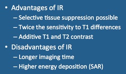
Inversion recovery techniques provide unique image contrasts in three important ways: 1) ability to selectively suppress ("null") the signal from any given tissue based on its T1-value; 2) superior discrimination of tissues based on T1 relaxation times; 3) additive (rather than competitive) T1 and T2 effects.
Selective Tissue Suppression
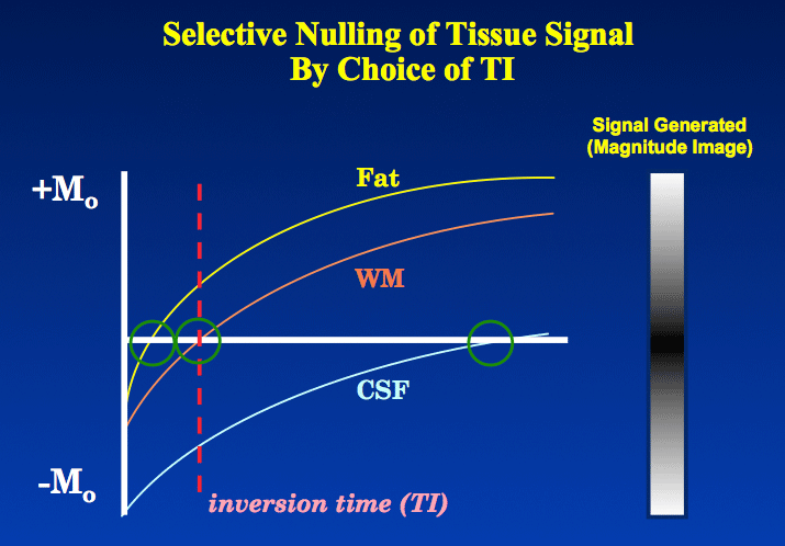
After the 180°-inverting pulse has been applied, the initial longitudinal magnetization (Mz) of tissues has been flipped into the −z-direction (opposite to Bo). During the inversion time (TI) interval (between this first 180° and 90° pulses), the magnetization of the inverted tissues regrows under T1 relaxation toward the +z-direction. During this process a time occurs where the Mz of each tissue passes through zero. If TI is set to that particular value (and signal generation initiated at that point), the tissue passing through zero will produce no signal and be effectively suppressed or "nulled". For most tissues the null point typically occurs when TI ≈ 0.69 x T1.
After the 180°-inverting pulse has been applied, the initial longitudinal magnetization (Mz) of tissues has been flipped into the −z-direction (opposite to Bo). During the inversion time (TI) interval (between this first 180° and 90° pulses), the magnetization of the inverted tissues regrows under T1 relaxation toward the +z-direction. During this process a time occurs where the Mz of each tissue passes through zero. If TI is set to that particular value (and signal generation initiated at that point), the tissue passing through zero will produce no signal and be effectively suppressed or "nulled". For most tissues the null point typically occurs when TI ≈ 0.69 x T1.
|
Up to Twice the Dynamic Range
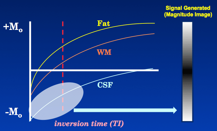
Because of the 180° inverting pulse, tissues in IR undergo T1 relaxation up to twice the dynamic range compared to SE. This means that IR can potentially discriminate among tissues based on subtle differences in T1 characteristics better than SE. This feature is illustrated in the graphs to the right where SE and IR are compared. |
|
Additive T1 and T2 contrast
Many types of pathology (eg, tumors, infarctions, infections) have elevated T1 and T2 values resulting from increased free water content compared to background tissue. In routine SE imaging ↑T1 generally results in decreased signal intensity while ↑T2 results in increased intensity. In other words, the combined T1 and T2 effects compete with and partially cancel each other in SE imaging. |
In IR imaging, however, especially using short to medium TI values, an ↑T1 will result in increased signal intensity that adds to the postive signal effects due to ↑T2. The reason for this is that when TI is less than that at the null point (< 0.69 x T1), the magnetization of the long T1 pathology will remain inverted and produce a high signal on magnitude reconstructed IR images.
Disadvantages of IR
In spite of all these unique features and advantages, there are several disadvantages for IR compared to SE and GRE techniques:
In spite of all these unique features and advantages, there are several disadvantages for IR compared to SE and GRE techniques:
- Longer scan times
- Increase in flow-related artifacts
- Signal-to-noise can decrease as tissues are suppressed
- Higher specific absorption rate (SAR) due to additional 180° pulses
Advanced Discussion (show/hide)»
No supplementary material yet. Check back soon!
References
Bydder GM, Young IR. MR imaging: clinical use of the inversion recovery sequence. J Comput Assist Tomogr 1985; 9:659-675.
Bydder GM, Young IR. MR imaging: clinical use of the inversion recovery sequence. J Comput Assist Tomogr 1985; 9:659-675.
Related Questions
What is the inversion recovery pulse sequence?
What is the inversion recovery pulse sequence?


