|
Caipirinha is Brazil's national cocktail, made from sugar cane liqueur (cachaça), sugar, ice, and lime. It received increased world-wide recognition as the result of news coverage of the 2014 FIFA World Cup.
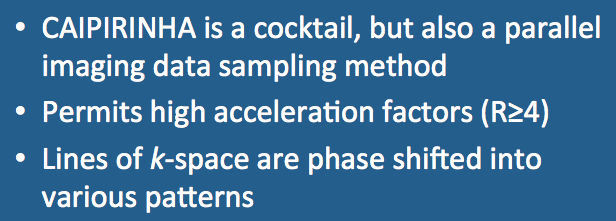
In MRI, however, CAIPIRINHA is a parallel imaging technique offered by Siemens and used primarily for 3D breath-hold abdominal imaging. The acronym stands for Controlled Aliasing in Parallel Imaging Results in Higher Acceleration.
|
Both SENSE and GRAPPA can be performed in 3D acquisition mode using acceleration in two phase-encoding directions simultaneously. Due to variability in coil geometries and sensitivities, however, severe residual aliasing artifacts and amplified noise may occur when high R-values are chosen.
CAIPIRINHA uses a group unique k-space sampling patterns to reduce pixel aliasing and overlap on reconstructed images. Acquired points in k-space are shifted from one another by applying additional offsets to the phase-encoding gradient tables.
CAIPIRINHA uses a group unique k-space sampling patterns to reduce pixel aliasing and overlap on reconstructed images. Acquired points in k-space are shifted from one another by applying additional offsets to the phase-encoding gradient tables.
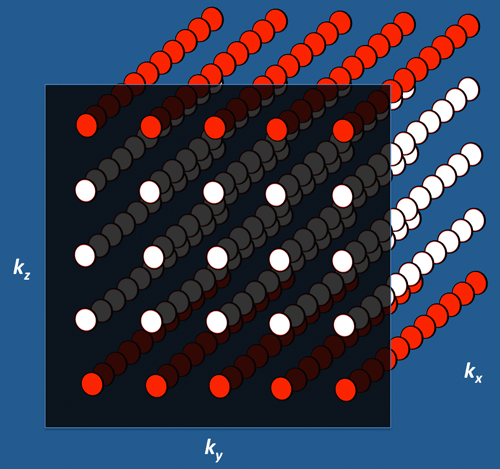
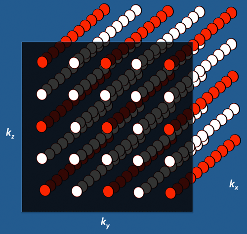
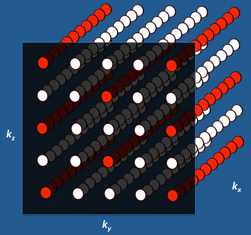
Below are three possible k-space filling patterns for a GRAPPA-like parallel imaging sequence with R=4. Note that the readout direction (kx) is directed into the page. The red circles represent acquired data points; the white circles represent missing data points that must be estimated from the known (red) points during reconstruction.
In the first diagram acceleration has been performed along a single direction (kz) with every 4th line of data acquired. This strategy often leads to increased noise and residual aliasing artifacts.
In the second diagram a better strategy has been adopted with the acceleration evenly split between the ky and kz directions. Every other line of data has thus been skipped in each of the two phase-encode directions. The total acceleration remains at 4 (R = 2 x 2).
In the second diagram a better strategy has been adopted with the acceleration evenly split between the ky and kz directions. Every other line of data has thus been skipped in each of the two phase-encode directions. The total acceleration remains at 4 (R = 2 x 2).
The third diagram shows a CAIPIRINHA pattern of sampling with R=2 in each phase-encode direction plus a phase shift of data sampling in the middle column (and every 4th column thereafter). In other words, the k-space sampling pattern can be viewed as staggered or sheared. This phase offset improves the accuracy of reconstruction while reducing noise and aliasing. Aliasing artifacts are still present but they are shifted to the corners of image space and are less likely to become concentrated in a few locations.
At present one of the most common uses of CAIPIRINHA is for liver studies where highly accelerated, excellent quality images can be obtained during passage of contrast. A clinical example is shown below. Other uses of CAIPIRINHA include diffusion-weighted imaging and MRA.
CAIPIRINHA may be operated in multislice mode (MS-CAIPIRINHA), 2D-mode (2D-CAIPIRINHA), or in a 3D pattern that takes advantage of coil sensitivity profiles along the readout direction (Wave-CAIPI). (Note that most other 3D PI techniques only exploit coil sensitivity variations along the phase-encoding and partition-encoding axes and ignore variations along the readout-axis.) As such, Wave-CAIPI can boast acceleration factors in the range of 9-12 with low artifacts and negligible g-factor penalties, finding great utility in high-resolution volumetric imaging.
Advanced Discussion (show/hide)»
Additional Notes on CAIPIRINHA
Although an example using GRAPPA was shown above, CAIPIRINHA can be used with either SENSE or GRAPPA algorithms. It may also be used with non-cartesian methods.
In addition to the so-called Δ=2 alternating phase method shown in the third diagram above, many different CAIPIRINHA sampling patterns are possible. The number of possibilities exponentially increases with the acceleration factor (R).
References
Caipirinha. Wikipedia, the Free Encyclopedia.
Bilgic B, Gagoski BA, Cauley SF, et al. Wave-CAIPI for highly accelerated 3D imaging. Magn Reson Med 2015; 73:2152-62. [DOI LINK]
Breuer F, Blaimer M, Griswold M, Jakob P. Controlled aliasing in parallel imaging results in higher acceleration (CAIPIRINHA). MAGNETOM Flash 2012;1:2-7. (Siemens promotional brochure).
Breuer FA, Blaimer M, Mueller MF, et al. Controlled aliasing in volumetric parallel imaging (2D CAIPIRINHA). Magn Reson Med 2006; 55:549-556.
Hamilton J, Franson D, Seiberlich N. Recent advances in parallel imaging for MRI. Prog NMR Spectr 2017; 101:71-95. [DOI LINK]
Polak D, Cauley S, Bilgic B, et al. Ultrafast multi-contrast high-resolution 3D brain MRI: a technical description of Wave-CAIPI. MAGNETOM Flash 2020; 76:2-5. [DIRECT LINK]
Yutzy SR, Seiberlich N, Duerk JL, Griswold MA. Improvements in multislice parallel imaging using radial CAIPIRINHA. Magn Reson Med 2011; 65:1630-1637.
Related Questions
What is parallel imaging? How does this differ from "regular" imaging?
What is parallel imaging? How does this differ from "regular" imaging?


