|
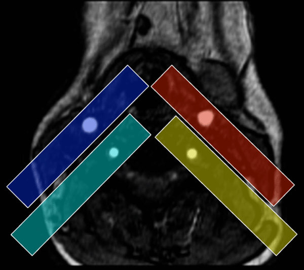
The technique you are describing is known as region-selective ASL. Instead of using a single large slab inversion pulse, a set of thinner pulses are individually played out over each of the major feeding arteries to an organ. This allows the separate perfusion contribution of each vessel on an anatomic basis to be measured. In the example shown, individual inversion pulses are applied in the neck over the L&R carotid arteries (red and blue) and the L&R vertebral arteries (yellow and green). This case shows that the left carotid and right vertebral arteries each supply disproportionate portions of the anterior and posterior cerebral circulations respectively.
|
Advanced Discussion (show/hide)»
No supplementary material yet. Check back soon!
References
Davies NP, Jezzard P. Selective arterial spin labeling (SASL): perfusion territory mapping of selected feeding arteries tagged using two-dimensional radiofrequency pulses. Magn Reson Med 2003; 49:1133-1142.
van Laar JP, van der Grond J, Hendrikse J. Brain perfusion territory imaging: methods adn clinical applications of selective arterial spin-labeling MR imaging. Radiology 2008; 246:354-364
Related Questions
Can you briefly explain the difference between the various ASL methods? Which is the best?
Can you briefly explain the difference between the various ASL methods? Which is the best?


