 MR image of the brain (left) and ¹H MR spectrum from the large
yellow voxel. The location of each spectral peak along the horizontal
chemical shift (δ) axis allows identification of each metabolite, while
the area under each peak is roughly proportional to the
metabolite's concentration.
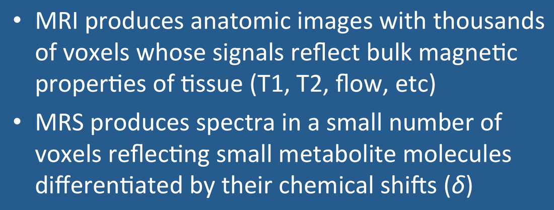
As its name implies, MR Imaging (MRI) is primarily concerned with the production of anatomic images. MRI scans usually contain many thousands of voxels whose signals reflect the bulk magnetic properties (e.g., T1, T2, susceptibility, flow) of the tissues they contain. The signals of interest recorded by MRI come predominantly from protons in water and fat.
By comparison, the goal of MR Spectroscopy (MRS) is to analyze the chemical composition of tissues in a very small number of much larger voxels. By excluding the overwhelming signals from water and fat, MRS can detect small metabolites existing in millimolar (mM) concentrations. These metabolites can be differentiated because they resonate at slightly different frequencies based on their local chemical environments. The degree of frequency separation between two molecular species is characterized by their chemical shift (δ), a small number displayed on the horizontal axis below their spectra. The relative areas under each peak are roughly proportional to the number of nuclei in that particular chemical environment.
By comparison, the goal of MR Spectroscopy (MRS) is to analyze the chemical composition of tissues in a very small number of much larger voxels. By excluding the overwhelming signals from water and fat, MRS can detect small metabolites existing in millimolar (mM) concentrations. These metabolites can be differentiated because they resonate at slightly different frequencies based on their local chemical environments. The degree of frequency separation between two molecular species is characterized by their chemical shift (δ), a small number displayed on the horizontal axis below their spectra. The relative areas under each peak are roughly proportional to the number of nuclei in that particular chemical environment.
Advanced Discussion (show/hide)»
No supplementary material yet. Check back soon!
References
Arnold JT, Dharmati S, Packard ME. Chemical effects on nuclear induction signals from organic compounds. J Chem Phys 1951; 19:507 (See the world's first MR spectrum recorded — ethanol).
Cresshull ID, Gordon RE, Hanley PE, et al. Localization of metabolites in animal and human tissue using ³¹P topical magnetic resonance. Bull Magnet Reson 1980; 2:426. (First MRS spectrum recorded in a human forearm.)
Proctor WG, Yu FC. The dependence of a nuclear magnetic resonance frequency upon chemical compound. Phys Rev 1950; 77:717. (This is the famous paper where changes in resonance frequency — later to be known as "chemical shifts" — were first reported among several nitrogen compounds, the basis for NMR spectroscopy).
Ross BD, Radda GK, Gadian DG, et al. Examination of a case of suspected McArdle's syndrome by ³¹P nuclear magnetic resonance. N Engl J Med 1981; 304:1338-1342. (First application of MRS to a human disease.)
Related Questions
What is meant by a chemical shift?
How are proton chemical shifts measured on the δ scale?
What is meant by a chemical shift?
How are proton chemical shifts measured on the δ scale?
