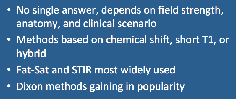
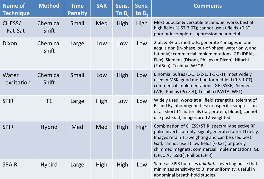
Yes, a number of different methods exist for suppressing high signal from fat to better visualize pathology or contrast enhancement. No single answer exists as to the "best" technique; the choice of technique depends on field strength, body part imaged, presence of gadolinium, proximity of metallic hardware, and clinical scenario.
As an overview, water-fat separation methods are based on: 1) the water-fat chemical shift (resonance frequency difference); 2) the short T1 of fat; or 3) both (hybrid techniques).
Chemical-shift Techniques
In-Phase/Out-of-Phase Imaging. This is not really a fat-suppression technique, but is simple and straightforward method to characterize tissues based on differences in water and fat resonance frequencies. By slight adjustment of the echo time (TE), signals from water and fat protons in the same voxel can be made to constructively or destructively interfere. This method is used primarily in abdominal imaging to characterize certain diseases (adrenal adenomas, hepatic steatosis).
Dixon Technique(s). A group of techniques closely related to the qualitative in-phase/out-of-phase imaging methods described above. Acquiring 2, 3 or more echoes at different TE's, "water only" and "fat only" images can be extracted through sophisticated mathematical processing. Fat quantification is also possible. The Dixon technique is widely used in abdominal imaging, imaging of the extremities, and the spine. It is less sensitive to Bo and B1 heterogeneity than CHESS/Fat-Sat methods.
CHESS (Fat-Sat). This is the most widely used method for fat suppression. An RF-pulse tuned to the fat resonance frequency together with a spoiler gradient saturates and dephases fat protons, leaving only water protons to produce a signal. The technique is very versatile and can be appended to any pulse sequence. It works best at 1.0T and higher and cannot be used below 0.3T (due to close position of spectral peaks). It requires a homogeneous field to work properly and thus may fail around metallic hardware, imaging far from isocenter, or in anatomic regions with susceptibility distortions (sinuses, head and neck).
Water Excitation. Instead of suppressing fat, these techniques use a short series of RF pulses to selectively excite water protons. No spoilers are needed. To date, water excitation methods have been used primarily in the musculoskeletal system, especially for evaluation of cartilage. Some 3D applications in the breast and liver have also been reported.
Technique Based on Short T1 of Fat
Short TI Inversion Recovery (STIR). This is another widely used sequence applicable at all field strengths. Because fat has a shorter T1 than nearly all other materials in the body, its signal can be selectively nulled using a magnitude-reconstructed inversion recovery sequence with short TI values (150-180 msec at 1.5T). The method is relatively insensitive to field inhomogeneities and can be used near metal and over large fields-of-view. The signal suppression is not specific to fat; any short T1 material (e.g., melanin, methemoglobin, mucus) will be nulled. Thus STIR cannot be used post-gadolinium to demonstrate contrast-enhancement, its most important limitation.
Hybrid Techniques
SPIR and SPAIR. These techniques combine a greater than 90º chemical-shift selective (CHESS) pulse with an inversion delay and spoiling to null the signal from fat. The main difference between SPIR (Spectral Presaturation with Inversion Recovery) and SPAIR (Spectral Attenuated Inversion Recovery) is that the latter uses a full 180º adiabatic inverting pulse that is insensitive to B1 inhomogeneity. Both methods are sensitive to Bo inhomogeneities.
Advanced Discussion (show/hide)»
No supplementary material yet. Check back soon!
References
Bley TA, Wieben O, François CJ, et al. Fat and water magnetic resonance imaging. J Magn Reson Imaging 2010; 31:4-18. (good review) [DOI Link]
Del Grande F, Santini F, Herzka DA, et al. Fat-suppression techniques for 3-T MR imaging of the musculoskeletal system. RadioGraphics 2014; 34:217-233. (Good recent review, focused on the musculoskeletal system).
Horger W. Fat suppression in the abdomen. MAGNETOM Flash 2007;3:114-119. (Good overview of multiple techniques, obviously focused exclusively on Siemens' products).
Related Questions
What makes fat and water behave so differently on MRI?
What makes fat and water behave so differently on MRI?

