Parallel imaging is a widely used technique where the known placement and sensitivities of receiver coils are used to assist spatial localization of the MR signal. Having this additional information about the coils allows reduction in number of phase-encoding steps during image acquisition. This, in turn, potentially results in a several-fold reduction in imaging time.
Although the details of how coil-specific data is translated into image information is somewhat complex, the basic idea underlying parallel imaging should not be. In fact, the core principle of parallel imaging is actually more easily grasped by most students than frequency- or phase-encoding.
|
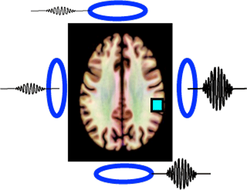
Consider, for example, a set of 4 receiver coils surrounding a head being used to detect the MR signal arising from a pixel as shown in the diagram left. Because each coil is at a different distance from the pixel, the signal recorded by each coil varies as a function of position (closer coils have stronger signals). Just by looking at the relative intensities from each coil, it is possible to make a crude prediction of the approximate source of the MR signal. Even without using phases, frequencies, or Fourier Transforms, we already know the signal is coming from somewhere in the left parietal lobe!
|
In "regular" (non-parallel) imaging, multiple surface coils may be used to detect the MR signal, but their individual outputs are combined into one aggregate complex signal that is digitized and processed into the final image. In parallel imaging, conversely, the signals from individual coils are amplified, digitized, and processed simultaneously "in parallel" along separate channels, retaining their identities until near the end.
|
Parallel imaging has become practical in modern MRI because of advancements in telecommunications technology that have significantly reduced the cost of RF-digital processing systems. Today MR vendors offer commercial systems able to support 200+ coils and 128+ receiver channels.
|
Advanced Discussion (show/hide)»
No supplementary material yet. Check back soon!
References
Blaimer M, Breuer F, Mueller M, Heidemann RM, Griswold MA, Jakob PM. SMASH, SENSE, PILS, GRAPPA. How to choose the optimal method. Top Magn Reson Imaging 2004;15:223-236 [review].
Deshmane A, Gulani V, Griswold MA, Seiberlich N. Parallel MR imaging. J Magn Reson Imaging 2012;36:55-72. (review)
Glockner JF, Hu HH, Stanley DW, et al. Parallel MR imaging: a user's guide. Radiographics 2005;25:1279-1297.
Hamilton J, Franson D, Seiberlich N. Recent advances in parallel imaging for MRI. Prog NMR Spectr 2017; 101:71-95. [DOI LINK]
Larkman DJ, Nunes RG. Parallel magnetic resonance imaging. Phys Med Biol 2007;52:R15-R55 [review]
Yanasak N, Clarke G, Stafford RJ et al. Parallel Imaging in MRI: Technology, applications, and quality control. American Association of Physicists in Medicine. Report No 118. June, 2015.
Related Questions
How does the scanner know the locations of all the MR signals?
Is PI a special type of pulse sequence? Can it be performed with any coil in any direction?
How does the scanner know the locations of all the MR signals?
Is PI a special type of pulse sequence? Can it be performed with any coil in any direction?


