|
Stents are widely used throughout the entire arterial system, ranging from vessels several centimeters across (like the thoracic aorta) down to vessels only a few millimeters in diameter (like coronary and intracranial arteries). Simple stents are cylindrical solid or mesh-like tubes made of metal or plastic whose function is to maintain vascular patency in the presence of stenosis or compression. A stent-graft is an expandable metal stent covered with fabric that forms its own lumen and is commonly placed in aneurysms. Nearly all metal stents are made of non-ferric materials such as 300-series stainless steel, Nitinol, Elgiloy, or other alloys. Non-covered stents may be bare (metal only) or impregnated with a medication (so-called drug-eluting stents).
|
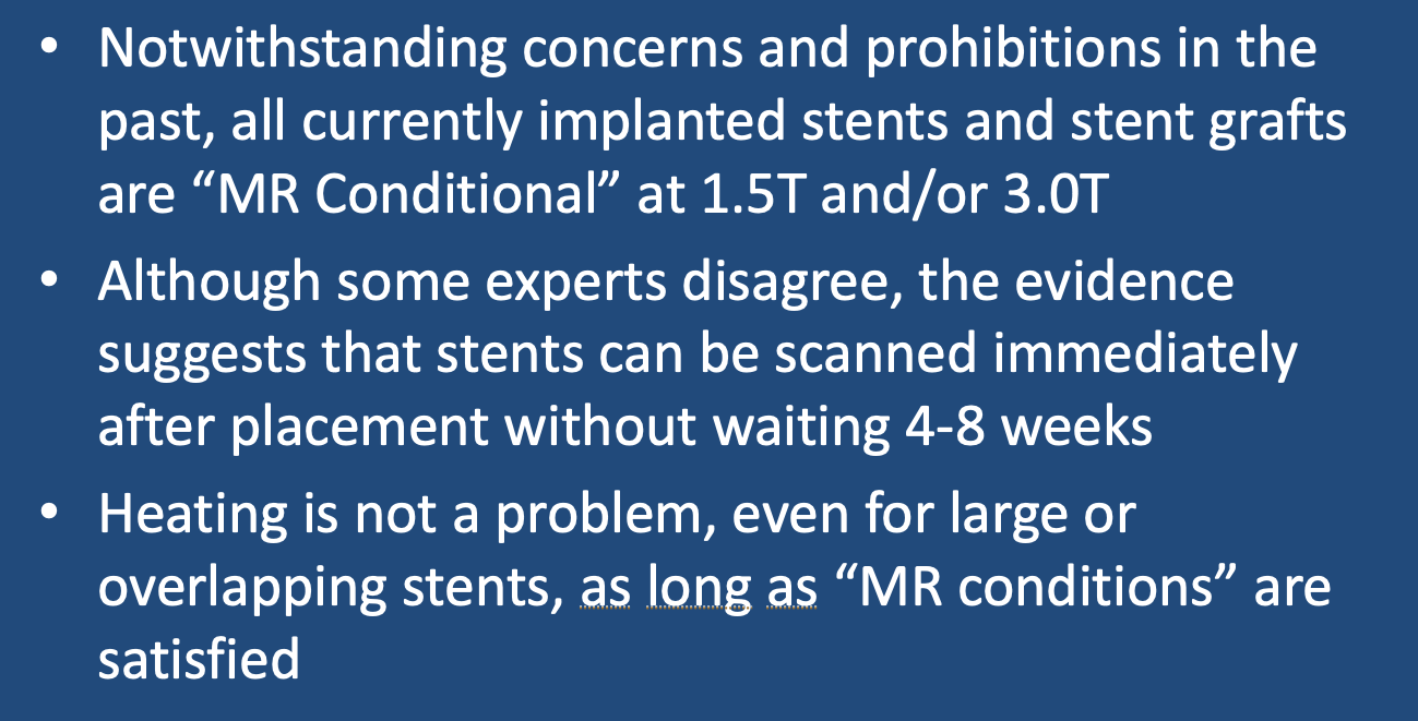
To my knowledge, there are no currently implanted stents that are considered MR Unsafe. In the past the Zenith AAA stainless steel stent-graft was placed in this category by its manufacturer (Cook), but this restriction has now been lifted. Like other metallic stents it is considered MR Conditional at 1.5T and 3.0T. Although safe to scan as long as conditions are followed, some stents like the Zenith produce a substantial susceptibility artifacts making assessment of stent patency by MRI difficult.
Another outdated concept is that one must wait 4-8 weeks before scanning a patient with a newly-implanted metal stent. The idea was that the stent needed time to "settle in" and become incorporated in the vessel wall before risking displacement by magnetic forces. As recently as 10 years ago, the package inserts of many stents, especially uncovered coronary stents, carried a warning not to scan patients in the first 6 weeks unless absolutely necessary. Since that time, several papers have been published demonstrating the safety of scanning patients immediately after stent placement; so that is the protocol I follow and recommend.
Metallic stents may indeed undergo heating during RF-excitation, but this also does not seem to be a major problem even with overlapping stents or with big aortic stent-grafts, in part because flowing blood serves to diffuse away whatever heat is locally generated. The "conditions" associated with some stents recommend that whole-body-averaged SAR levels not exceed 2 W/kg and a maximum of 15 minutes per sequences, while other stents permit up to 4 W/kg. If you don't know the exact model of the stent you are scanning it is therefore safer to use the lower limit.
Advanced Discussion (show/hide)»
To be fair and balanced, there is a single controversial case report from 2013 (Parthasarathy H, Saeed O, Marcuzzi D, Cheema AN. MRI-induced stent dislodgment soon after left main coronary artery stenting. Circ Cardiovasc Interv. 2013;6:e58–e59) wherein a very short left main coronary stent perched at the ostium was found displaced to an iliac artery after a 1.5T MRI was performed 10 days after placement. Although the authors blamed the MRI for the displacement, subsequent letters to the editor cast doubt on this assertion, providing evidence that the original stent was poorly chosen and placed and that spontaneous displacement of similarly placed stents had been reported in the absence of MRI.
References
Levine GN, Gomes AS, Arai AE, et al. Safety of magnetic resonance imaging in patients with cardiovascular devices: An American Heart Association scientific statement from the Committee on Diagnostic and Interventional Cardiac Catheterization, Council on Clinical Cardiology, and the Council on Cardiovascular Radiology and Intervention: Endorsed by the American College of Cardiology Foundation, the North American Society for Cardiac Imaging, and the Society for Cardiovascular Magnetic Resonance. Circulation 2007; 116:2878–2891.
Maralani PJ, Schieda N, Hecht EM, et al. MRI safety and devices: an update and expert consensus. J Magn Reson Imaging 2019; 51:657-674. [DOI LINK]
Porto I, Selvanayagam J, Ashar V, Neubauer S, Banning AP. Safety of magnetic resonance imaging one to three days after bare metal and drug-eluting stent implantation. Am J Cardiol 2005; 96:366-8. [DOI LINK]
Shellock FG, Shellock VJ. Metallic stents: Evaluation of MR imaging safety. AJR Am J Roentgenol 1999;173:543–547.
Syed MA, Carlson K, Murphy M, et al. Long-term safety of cardiac magnetic resonance imaging performed in the first few days after bare-metal stent implantation. J Magn Reson Imaging 2006; 24:1056–1061.
Related Questions
How about other GU devices like nephrostomy tubes and stents?
Is there an increased risk of IVC filters moving during MRI?
What do you do about tracheobronchial airway devices like stents, valves and coils?
How about other GU devices like nephrostomy tubes and stents?
Is there an increased risk of IVC filters moving during MRI?
What do you do about tracheobronchial airway devices like stents, valves and coils?

