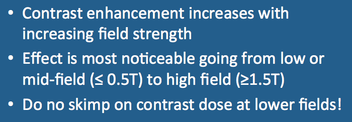
The degree of contrast enhancement (for a given dose, agent, and pulse sequence) increases with increasing field strength, being most noticeable between low-field (≤0.5T) and high-field (≥1.5T) imaging. To understand why this occurs we must review several basic concepts of relaxation and relaxivity introduced in a prior Q&A.
The observed T1 relaxation rate (1/T1obs) caused by contrast material accumulating in a tissue is given by
1/T1obs = 1/T1t + r1 • [C]
where 1/T1t is the base relaxation rate of the tissue without contrast, [C] is the concentration of contrast agent within the tissue, and r1 is the relaxivity of the contrast agent. By algebraic rearrangement it is possible to derive an expression for the percent change in tissue relaxation due to the contrast agent:
%Δ(1/T1obs) = (1/T1obs − 1/T1t ) ÷ (1/T1t) = T1t • r1 • [C]
For a T1-weighted pulse sequence, percent change in 1/T1obs can be taken as a surrogate for relative lesion enhancement. This equation is consistent with several features about contrast enhancement well recognized by clinical radiologists:
- Contrast enhancement is more obvious in tissues with baseline long T1t values (i.e., tissues that are dark on T1-weighted images).
- More contrast [C] in a lesion means more enhancement.
- Contrast agents with higher T1 relaxivities (r1) values produce stronger enhancement.
To understand how the field-strength dependence of contrast enhancement, we must consider how T1t and r1 each change as a function of Bo.
The field-dependence of tissue relaxation times has been discussed in a prior Q&A. In general, tissue T1 values increase with increasing field strength. This phenomenon occurs because the Larmor frequency scales with field strength and T1 relaxation requires energy exchanges near the Larmor frequency to be effective. With increasing Larmor frequencies a progressive mismatch occurs with molecular motions in solid tissues, resulting in inefficient energy transfer and longer T1 values.
|
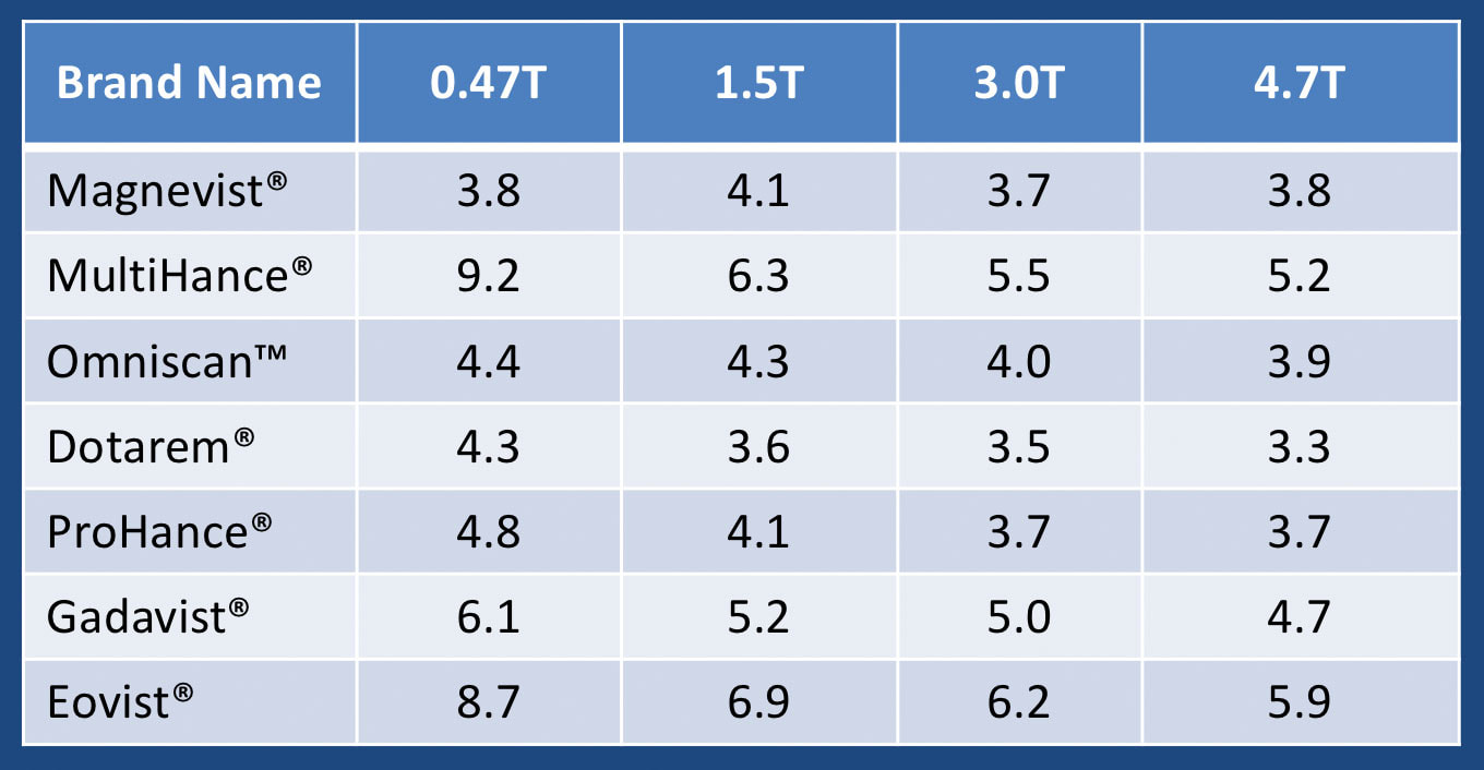
The field dependence of gadolinium-based MR contrast agents depends on the range of field strengths considered as well as the particular agent. The table right shows T1 relaxivities for nine commercially available contrast agents.
In general a slight decrease in r1 is noted for all agents with increasing field strength. The greatest percent decrease is seen from between low-field (≤0.5T) and high-field (≥1.5T). |
The largest field strength dependency is noted for contrast agents with higher degrees of protein binding (MultiHance®, Eovist®). This results from the accentuated Larmor frequency-molecular motion mismatch similar to that seen with solid tissues described above.
Because tissue T1 values increase while contrast agent relaxivities are about the same or decrease only slightly with increasing field strength, the net effect is an increase in relative contrast enhancement as field strength increases.
Advanced Discussion (show/hide)»
No supplementary material yet. Check back soon!
References
Bottomley PA, Foster TH, Argersinger RE, Pfeifer LM. A review of normal tissue hydrogen relaxation times and relaxation mechanisms from 1-100 MHz: dependence on tissue type, NMR frequency, temperature, species, excision, and age. Med Phys 1984;11:425-448.
Elster AD. Field-strength dependence of gadolinium enhancement: theory and implications. AJNR Am J Neuroradiol 1994; 15:1420-1423.
Hagberg GE, Scheffler K. Effect of r1 and r2 relaxivity of gadolinium-based contrast agents on the T1-weighted MR signal at increasing magnetic field strengths. Contr Media Mol Imag 2013; 8:456-465.
Rinck PA, Muller RN. Field strength and dose dependence of contrast enhancement by gadolinium- based MR contrast agents. Eur Radiol 1999;9:998-1004
Rohrer M, Bauer H, Mintorovitch J et al. Comparison of magnetic properties of MRI contrast media solutions a different magnetic field strengths. Invest Radiol 2005; 40:715-724.
Related Questions
How do T1 and T2 values vary as a function of field strength?
How do T1 and T2 values vary as a function of field strength?

