|
During the first half of the 20th Century Isador Rabi, Felix Bloch, and Edward Purcell described the NMR phenomenon; but it was the next generation of scientists in the 1970's who discovered how to create images from the NMR signal. Primary credit (and the Nobel Prize) for MR imaging was awarded to Sir Peter Mansfield and Paul Lauterbur. However, Raymond Damadian must also be recognized for his innovation and construction of the first full-body human MR scanner.
|
Lauterbur's Contribution: Projectional NMR Tomography
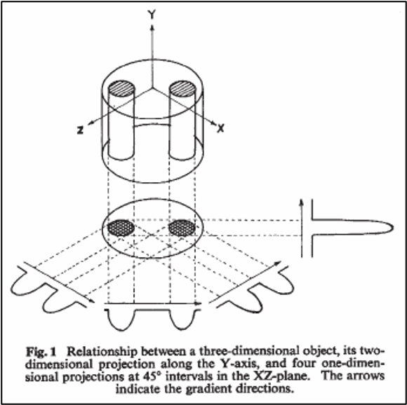
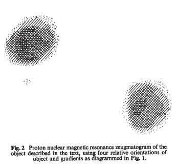
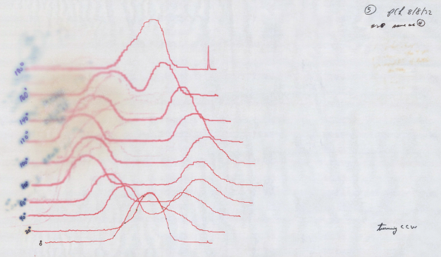
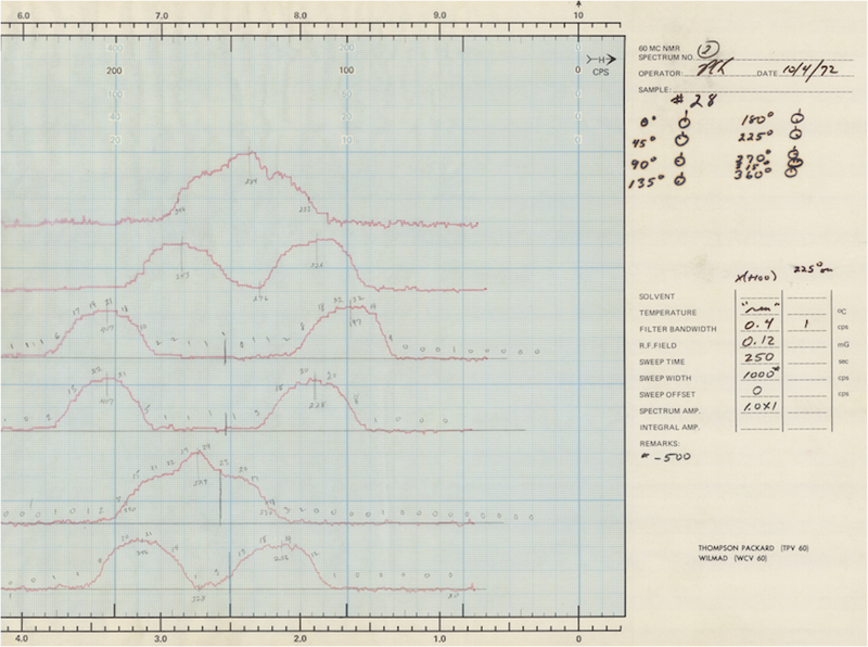
Paul Lauterbur (1909-2007), a chemist working at the State University of New York at Stony Brook, published the first true MR image in Nature in March, 1973. His experimental setup involved two 1-mm-diameter tubes filled with water placed in an 1.4T magnet. Applying magnetic field gradients rotated successively by 45°, he was able to obtain four different 1-dimensional projections of the NMR signal. These data were then mathematically "back-projected" to form a 2-dimensional tomographic image. Because the result depended on the combined effects of two magnetic fields, Lauterbur named his technique "zeugmatography" after the Greek word, zeugma, meaning "that which is used for joining." Shortly thereafter, Lauterbur produced crude images of his first living subject: a tiny clam.
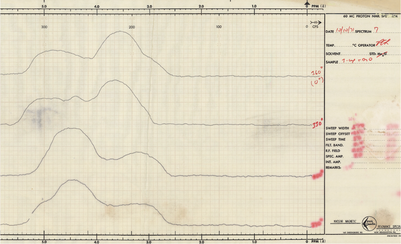
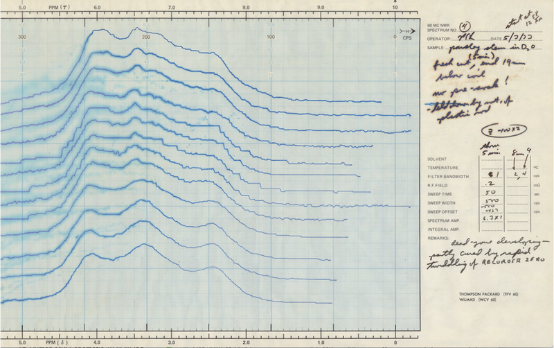
Figures 1 and 2 from Lauterbur's landmark paper in Nature (1973) and pages from his lab notebook (1971-1972) showing progress to the finished work. (Images courtesy of Andrew Webb through Joshua Shimony)
Mansfield's Contribution: Use of a field gradient for slice selection
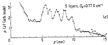 From Mansfield (1973). Five peaks corresponding
to five stacked blocks of solid camphor.
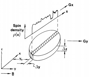
Also in 1973, Peter Mansfield (1933-2017), a physicist working at the University of Nottingham, demonstrated how a linear field gradient could be used to localize the NMR signal on a slice-by-slice basis. Mansfield's experimental setup involved stacking multiple 1-mm-thick sheets of solid camphor into the bore of an NMR spectrometer. Applying a magnetic field gradient perpendicular to the sheets, Mansfield measured the transient NMR signal response to an applied RF-pulse. Interference peaks similar to those seen in x-ray diffraction were observed, which when inverse Fourier transformed revealed discrete layers of the camphor sample.
Later in the decade, Mansfield and his collaborator, Andrew Maudsley, further refined this method into a line-scan technique, producing the first image of a human body part, a finger, in 1977.
Later in the decade, Mansfield and his collaborator, Andrew Maudsley, further refined this method into a line-scan technique, producing the first image of a human body part, a finger, in 1977.
Damadian's Contribution: Vision of a human-sized scanner to detect disease
|
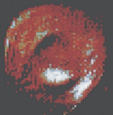
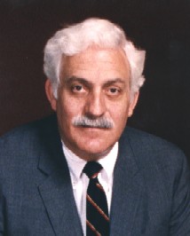
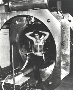
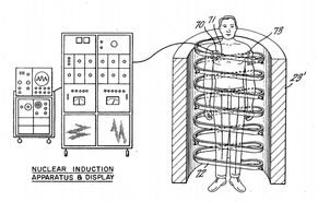
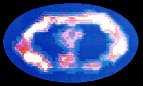
While Lauterbur and Mansfield were basic scientists, Raymond V. Damadian (b. 1936) was a physician, an Associate Professor of Medicine at the State University of New York - Brooklyn (Downstate). He looked at NMR from a different and original perspective — as a phenomenon that might be used to probe the body and diagnose human disease. In one of his landmark early papers (Science, 1971) Damadian demonstrated that cancer cells had longer T1 and T2 values than normal cells. In 1972 he filed a US patent application for an apparatus and method to detect cancer in tissue. Although the details of exactly how this 'apparatus" would produce images were not included in the application, Damadian and his team set out to build such a device which was named "Indomitable." By mid-summer, 1977, the first whole-body MR images were being produced, including the famous one shown below of his assistant's chest.
|
|
Damadian used a "sensitive point" method for spatial localization of the NMR signal. This was based on a saddle-shaped magnetic field where only a small volume at the center matched the resonance frequency of the RF pulse. The patient's body was physically moved in a rectangular pattern until signals from all pixels were obtained.
|
Damadian called his imaging method "field-focused NMR" or FONAR. This became the name of his company, the first to manufacture clinical MR scanners commercially. It was soon recognized that the field-focused method was far too slow and clumsy for routine clinical imaging, and so it was abandoned in favor of the methods of Lauterbur and Mansfield in subsequent versions of the scanner.
When the 2003 Nobel Prizes for Medicine were announced, Damadian considered it a personal injustice that he was excluded. He placed full-page ads in several large world newspapers urging the Nobel committee to change its mind. The decision stood.
When the 2003 Nobel Prizes for Medicine were announced, Damadian considered it a personal injustice that he was excluded. He placed full-page ads in several large world newspapers urging the Nobel committee to change its mind. The decision stood.
Advanced Discussion (show/hide)»
In addition to Damadian, Herman Carr had some claim to the Nobel Prize as well. In 1952 he showed how field gradients could be used to create 1-D images in an experimental device.
Below is a YouTube video about the contributions of Paul Lauterbur.
References
(Many papers, but all are worth at least perusing in that they reflect the early development of our field in the 1970's)
Damadian R. Tumor detection by nuclear magnetic resonance. Science 1971; 171:1151-3. (First measurements of T1 and T2 relaxations of a wide range of normal tissues, including several cancers that showed prolongation of T1 and T2)
Damadian RV. Apparatus and method for detecting cancer in tissue. US Patent # 3,789,832 (filed 3/17/1972, granted 2/5/1974).
Damadian R. Field focusing n.m.r (FONAR) and the formation of chemical images in man. Phil Trans R Soc Lond B 1980; 289:489-500.
Damadian R, Minkoff L, Goldsmith L et al. Field focusing nuclear magnetic resonance (FONAR): visualization of a tumor in a live animal. Science 1976; 194:1430-2.
Damadian R, Goldsmith M, Minkoff L. NMR in cancer: XVI. FONAR image of the live human body. Physiol Chem & Phys 1977; 9:97-109.
Garroway AN, Grannell PK, Mansfield P. Image formation in NMR by a selective irradiative process. J Phys C: Solid State Phys 1974; 7:L457-462.
Hinshaw WS. Image formation by nuclear magnetic resonance: the sensitive point method. J Appl Phys 1976; 47:3709-21.
Kumar A, Welti D, Ernst RR. NMR Fourier Zeugmatography. J Magn Reson 1975; 18:69-83.
Lauterbur PC. Image formation by induced local interactions: examples employing nuclear magnetic resonance. Nature 1973; 242:190-1.
Mansfield P. Multi-planar image formation using NMR spin echoes. J Phys C: Solid State Phys 1977; 10:L55-L58.
Mansfield P, Maudsley AA. Planar spin imaging by NMR. J Magn Reson 1977; 27:101-119.
Mansfield P, Grannell PK. NMR 'diffraction' in solids? J Phys C: Solid State Phys 1973; 6:L422-L426.
Mansfield P, Maudsley AA. Medical imaging by NMR. Br J Radiol 1977; 50:188-194. (First image of human finger)
|
© 2024 AD Elster, ELSTER LLC
All rights reserved. |
|


