|
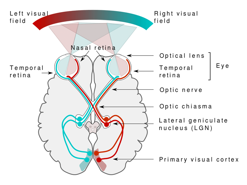
The basic anatomy of the visual pathway should be familiar to all neuroscience students: Light enters the eye, stimulating retinal cells, whose integrated output fibers travel along the optic nerves, partially crossing in the optic chiasm, and continuing along the optic tracts to synapse in the lateral geniculate nucleus (LGN) of the thalamus. Optic radiations emerge from the LGN to terminate on neurons in a specialized region of the occipital lobe — the primary visual cortex —also known as the striate cortex or visual area one (V1). Retinal projections to V1 are highly organized, with each visual field projecting to the contralateral cerebral hemisphere.
The most complex integration of visual information occurs in the extrastriate cortex, visual association areas denoted V2−V6 located in the adjacent occipital, temporal, and parietal lobes. Transmission to these areas occurs along two pathways: a ventral stream thought to be responsible for object recognition and the storage of long-term visual memories, and a dorsal stream associated with the spatial location of objects and perception of motion.
|
|
Historically, the earliest fMRI experiments from the 1990s involved activation of the visual cortex. Today the major clinical application of visual system fMRI is preoperative planning for patients with brain tumors, vascular malformations, and other lesions that impact the central visual pathways. DeYoe et al. have developed a set of paradigms based on slowly expanding checkerboard rings and wedges that produce a strong BOLD signal permitting both visual field eccentricity and angular position to be mapped in a reasonable time frame. The fMRI data acquired is often combined with diffusion tensor imaging to identify white matter tracts associated with these cortical areas.
|
Advanced Discussion (show/hide)»
A great deal of basic science research is currently being performed using fMRI to study the function of the visual association areas in humans. This has not yet reached the stage of clinical utility for presurgical planning, but may in the near future.
References
American Society of Functional Neuroradiology (ASFNR). Functional Imaging Paradigms (2007). Available from http://www.asfnr.org/wp-content/uploads/ASFNR-BOLD-Paradigms.pdf (visual paradigms on pp. 37-40)
DeYoe EA, Ulmer JL, Mueller WM, et al. Imaging of the functional and dysfunctional visual system. Semin Ultrasound CT MRI 2015; 36:234-248.
Drobyshevsky A, Baumann SB, Schneider W. A rapid fMRI task battery for mapping of visual, motor, cognitive and emotional function. NeuroImage 2006; 31:732-744. (A practical and fairly comprehensive protocol for eloquent cortex mapping that can be performed in 30 min)
Engel SA, Glover GH, Wandell BA. Retinotopic organization in human visual cortex and the spatial precision of functional MRI. Cereb Cortex 1997; 7:181-192. (Description of the traveling wave method, commonly used for visual fMRI experiments).
Mishkin M, Ungerleider LG, Macko KA. Object vision and spatial vision: two cortical pathways. Trends Neurosci 1983; 6:414-417. (famous paper describing the "two-stream hypothesis" for visual processing)
Warnking J, Dojat M, Guérin-Dungué A, et al. fMRI retinotopic mapping —step by step. NeuroImage 2002; 17:1665-1683.
Related Questions
Who invented functional MR imaging (fMRI)?
How are those activation "blobs" on an fMRI image created, and what exactly do they represent?
Who invented functional MR imaging (fMRI)?
How are those activation "blobs" on an fMRI image created, and what exactly do they represent?


