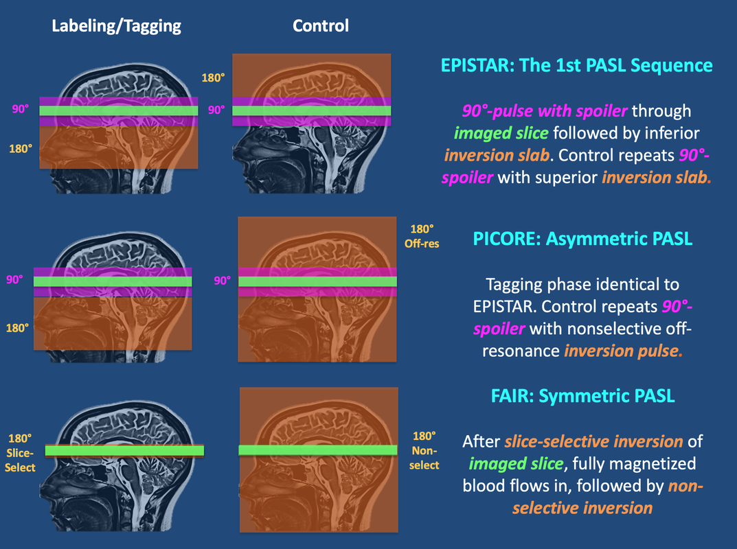
FAIR (Flow-sensitive Alternating Inversion Recovery) uses a slightly different pulsed labeling strategy that is symmetric with respect to the imaging slice. The tagged sequence begins with a spatially selective inversion pulse limited to a small region in and around the imaged slice. (The inversion pulse is usually slightly wider than the imaged slice to minimize artifacts from RF-side lobes and ensure uniform inversion). After this inversion pulse, "fresh" (fully magnetized) blood flows into the slice which serves as the "tag". This is followed by a non-slice-selective inversion pulse to the entire imaged volume as the "control". Versions of both PICORE and FAIR with Q2-TIPS bolus saturation (see Advanced Discussion) are currently offered as Siemens ASL products.
Advanced Discussion (show/hide)»
An additional difference between the three PASL techniques concerns the polarity of the signals between the tagged (St) and control (Sc) states. For EPISTAR and PICORE, inflowing arterial spins are inverted and thus St < Sc. For FAIR the tagged magnetization is not inverted while the control state is; hence St >Sc.
- Multi-slice STAR (Signal Targeting by Alternating Radiofrequency pulses) replaces the single distal inversion slab of the EPISTAR Control sequence with two proximal inversion slabs each at half amplitude applied in rapid succession. This has the same effect as CASL Solution #2 to create identical MT effects on the imaged slice but no net effect on flowing spins.
- PULSAR (PULsed StAR) uses the same inversion strategy as STAR but replaces the slice-selective 90°-pulse and spoiler gradient presaturation of the imaged slice using a more sophisticated cluster of 4 spectroscopic water-suppression pulses.
- QUASAR (QUantitative Star labeling of Arterial Regions) uses PULSAR labeling and control modules followed by a Look-Locker strategy sampling multiple time points for T1 estimation plus a repetitive Q2-TIPS-like saturation scheme for sharper definition of the arterial bolus. Other advanced features include perfusion quantification using an arterial input function (AIF) convolution method.
Both the leading and trailing edges of the bolus may be somewhat ill-defined due to transit delays and dispersion. Several pulse sequence modifications have been proposed to "sharpen" and thus improve edge definition of the bolus. One of the first was named QUIPSS (Quantitative Imaging of Perfusion using a Single Subtraction). QUIPSS involved adding an additional slab-select 90º, 18-lobe sinc saturation pulse just before readout in a PASL sequence applied either to the imaging volume (QUIPSS I) or labeling volume (QUIPSS II). A popular variant we use, Q2TIPS (QUIPSS II with Thin-slice TI1 Periodic Saturation), replaces this single thick-slab saturation pulse with a train of thin-slice saturation pulses at the distal end of the tagged region. Two operator-selectable parameters are available that determine the start and end times of this saturation train (and hence the edges of the bolus) Our Q2TIPS sequence uses 16 very selective suppression (VSS) pulses applied every 25 ms during a window 800-1200 ms after the initial inversion. These saturate a 2 cm slab of tissue leaving about a 1 cm gap proximal to the first imaging slice.
Edelman RR, Chen Q. EPISTAR MRI: Mulitslice mapping of cerebral blood flow. Magn Reson Med 1998; 40:800-805. (Later iteration of EPISTAR with modifications and multi-slice acquisition)
Edelman RR, Siewert B, Darby DG, et al. Qualitative mapping of cerebral blood flow and functional localization with echo-planar MR imaging and signal targeting with alternating frequency. Radiology 1994; 192:513-520. (one of the early descriptions of EPISTAR)
Golay X, Hendrikse J, Lim TCC. Perfusion imaging using arterial spin labeling. Top Magn Reson Imaing 2004; 15:10-27. (Good review plus a description of ASL of variants and acronyms such as FAIRER, TILT, BASE, and others).
Golay X, Peterson ET, Hui F. Pulsed Star labeling of arterial regions (PULSAR): a robust regional perfusion technique for high field imaging. Magn Res Med 2005; 53:15-21.
Kim S-G. Quantification of relative cerebral blood flow change by Flow-sensitive Alternating Inversion Recovery (FAIR) technique: application to functional mapping. Magn Reson Med 1995; 34:293-301. (original description of FAIR technique)
Luh W-M, Wong EC, Bandettini PA, Hyde JS. QUIPSS II with thin-slice TI1 periodic saturation: a method for improving accuracy of quantitative perfusion imaging using pulsed arterial spin labeling. Magn Reson Med 1999; 41:1246-1254 (description of the Q2TIPS method).
Petersen ET, Lim T, Golay X. Model-free arterial spin labeling quantification: approach for perfusion MRI. Magn Reson Med 2006; 55:219-232. (QUASAR method)
Wong EC, Buxton RB, Frank LR. Implementation of quantitative perfusion imaging techniques for functional brain mapping using pulsed arterial spin labeling. NMR in Biomed 1997; 10:237-249. (description of the PASL/PICORE method)
Can you briefly explain the difference between the various ASL methods? Which is the best?
What is pCASL and how does it differ from CASL and PASL?

