MR angiography is always evolving, with new methods and modifications of older methods being constantly proposed. Several new techniques have even made their ways into limited clinical practice, being offered by a small number of vendors as commercial products or work-in-progress (WIP) sequences for testing. Some of these newest and most promising MRA techniques are briefly described below.
|
Flow-Sensitive Dephasing (FSD)
FSD is a non-contrast subtraction technique similar to the 3D-gated FSE MRA method described in a prior Q&A. The FSD sequence simultaneously acquires a diastolic image (where both arteries and veins are bright) and a systolic image (where veins remain bright but arteries are dark). By subtracting these two images a pure arterial image remains.
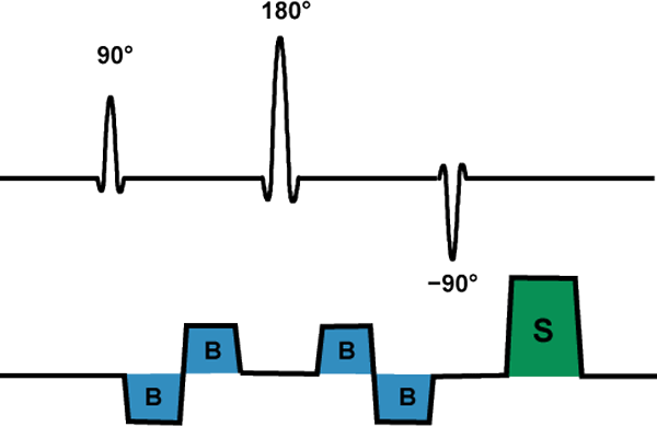
The FSD method is a T2-preparatory module that can be appended to either a 3D-balanced SSFP or 3D-FSE sequence. The FSD module consists of a cluster of three RF-pulses in a driven-equilibrium (DE) arrangement (90º/180º/−90º). The −90º flip-back pulse in the DE cluster helps restore longitudinal magnetization of blood, accentuating its signal. Sandwiched on either side of the 180º-pulse are mirror-image bipolar gradients. Unlike the bipolar gradients used in phase-contrast MRA, the gradients in FSD are turned on only during collection of the systolic image and are used to dephase arterial signal. A spoiler gradient is applied at the end of the module to prevent dephased spins from refocusing in later modules.
The choice of optimal gradient strength is tricky to preserve high arterial signal and prevent venous contamination. To detect flow in multiple directions, additional FSD modules performed in orthogonal orientations may be needed. Promising applications of FSD MRA include imaging the small vessels of the hands and feet.
|
|
Quiescent-Interval Single-Shot (QISS) MRA
|
QISS is a cardiac-gated, non-contrast inflow technique bearing some similarities to 2D time-of-flight (TOF) and inflow-enhanced SSFP MRA. It is especially designed for peripheral MRA. The sequence begins with a pair of closely-spaced 90º-RF pulses, one to saturate signal in the slice to be imaged and the other more distally located to suppress venous inflow. Next comes a quiescent interval (QI) of about 230 ms, during which fresh (unsaturated/fully magnetized) blood enters the imaging slice. A brief fat-suppression pulse is then applied to destroy any fat signal that has recovered during the QI. Finally, the desired arterial signal is acquired from the slice using a 2D single-shot, balanced SSFP sequence.
The long quiescent interval provides much more complete refreshment of blood than TOF methods with their much shorter TR's (≈ 30 msec). In theory this allows better detection of slowly flowing blood. Like 2D TOF, QISS performs best when arteries are long and perpendicular to the plane of imaging. Similar to stepped-table bolus-chase MRA, the peripheral arteries can be examined in 8-10 groups of 40-50 slices as the patient is moved through the magnet isocenter. Because SSFP readout is used, automatic reshimming may be needed at each station in the runoff.
|
Gated Time-of-Flight (TOF) Inflow MRA
Simple cardiac gating can improve the appearance of a 2D TOF MR angiogram at the expense of increased imaging time. Even further improvements can be achieved if the k-space ordering is adjusted so that the central lines of k-space occur during peak velocity (systolic phase). The primary advantage is the reduction of pulsatile flow artifacts causing physical movement of the artery and spatial blurring.
Philips offers a dual-gated method which acquires segmented data both in systole and diastole. This may offer some advantage in characterizing the degree of stenotic lesions.
Philips offers a dual-gated method which acquires segmented data both in systole and diastole. This may offer some advantage in characterizing the degree of stenotic lesions.
Advanced Discussion (show/hide)»
No supplementary material yet. Check back soon!
References
Edelman RR, Sheehan JJ, Dunkle E, et al. Quiescent-interval single-shot unenhanced magnetic resonance angiography of peripheral vascular disease: technical considerations and clinical feasibility. Magn Reson Med 2010; 63:951-958.
Fan Z, Saouaf R, Liu X, et al. Non-contrast MR angiography: flow-sensitive dephasing (FSD)-prepared 3D balanced SSFP. MAGNETOM Flash 2013; 4:2-7.
Fan Z, Sheehan J, Bi X, et al. 3D noncontrast MR angiography of the distal lower extremities using flow-sensitive dephasing (FSD)-prepared balanced SSFP. Magn Reson Med 2009; 62:1523-1532.
Lim RP, Fan Z, Chatterji M, et al. Comparison of nonenhanced MR angiographic subtraction techniques for infragenual arteries at 1.5T: a preliminary study. Radiology 2013; 267:293-304.
Hodnett PA, Koktzoglou I, Davarpanah AH, et al. Evaluation of peripheral arterial disease with nonenhanced quiescent-interval single-shot MR angiography. Radiology 2011; 260:282-293.
Related Questions
What is a driven equilibrium (fast recovery) pulse?
What is a driven equilibrium (fast recovery) pulse?


