|
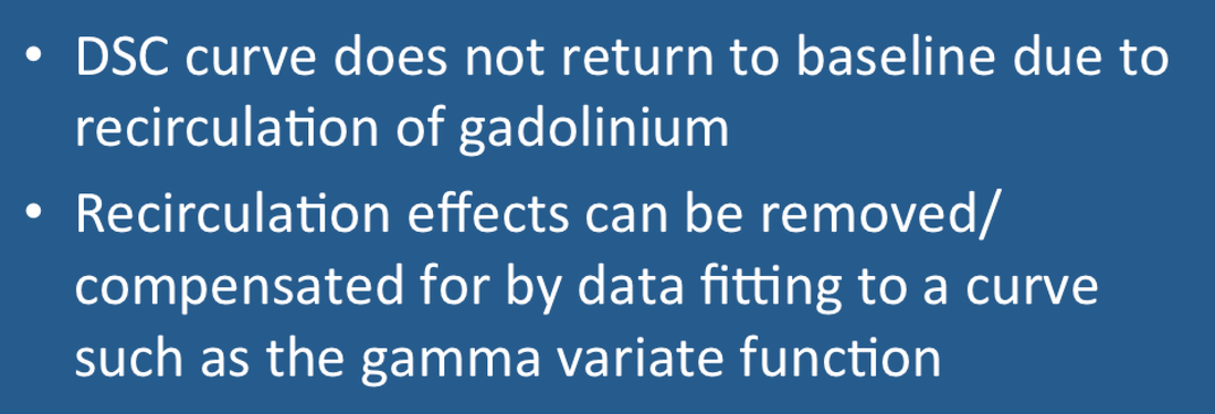
When gadolinium passes through tissue, T2/T2* dephasing occurs with a concomitant decrease in MR signal intensity. The dominant negative peak primarily reflects 1st pass of the initial bolus through the regional arterial circulation. Almost simultaneously, however, a second wave of gadolinium passes through the tissue that affects the size and timing of the first peak. This second wave represents recirculation of gadolinium-containing blood that has been shunted through the renal and coronary circulations and back into the heart. In addition to affecting the main peak, continued recirculation prevents the DSC signal from returning to its original baseline.
|
Advanced Discussion (show/hide)»
1. The gamma variate function can be written in several forms, but the one commonly used for first pass contrast agent experiments is given by the expression
S(t) = A(t−to)α e−(t−to)/β
where A is a scaling factor, to is the bolus arrival time, and α and β determine the shape of the distribution. To avoid recirculation effects being included in the gamma variate, often only the first portion of the contrast curve is used to estimate these parameters.
2. Whether and how to correct for recirculation effects is controversial. Paradoxically, blood volume estimates may be better when recirculation effects are not removed. Also, gamma variate fitting is inherently noisy as only the first portion of the uptake curve is sampled, thus generating problems of its own.
3. If a gamma variate method is selected, I recommend that a second output map (χ²-map) be generated to test for goodness of fit.
4. The typical DSC imaging sequence acquires data for a period of less than 30 seconds. In the setting of severe ischemia the first bolus of contrast may not reach the target organ during this period. Blood volume and other physiological parameters may therefore be underestimated.
Boxerman JL, Rosen BR, Weisskoff RM. Signal-to-noise analysis of cerebral blood volume maps from dynamic NMR imaging studies. J Magn Reson Imaging 1997; 7:528-37. (Disadvantages of gamma variate fitting).
Patil V, Johnson G. An improved model for describing the contrast bolus in perfusion MRI. Med Phys 2011; 38:6380-3. (An alternative to gamma variate fitting, the single compartment recirculation (SCR) model).
Thompson HK Jr, Starmer CF, Whalen RE, McIntosh HD. Indicator transit time considered as a gamma variate. Circ Res 1964; 14:502-515. (Original classic paper describing gamma variate fitting in indicator dilution experiments)
If gadolinium contrast is used to increase signal intensity on routine MR images, why does it decrease signal on DSC perfusion images?

