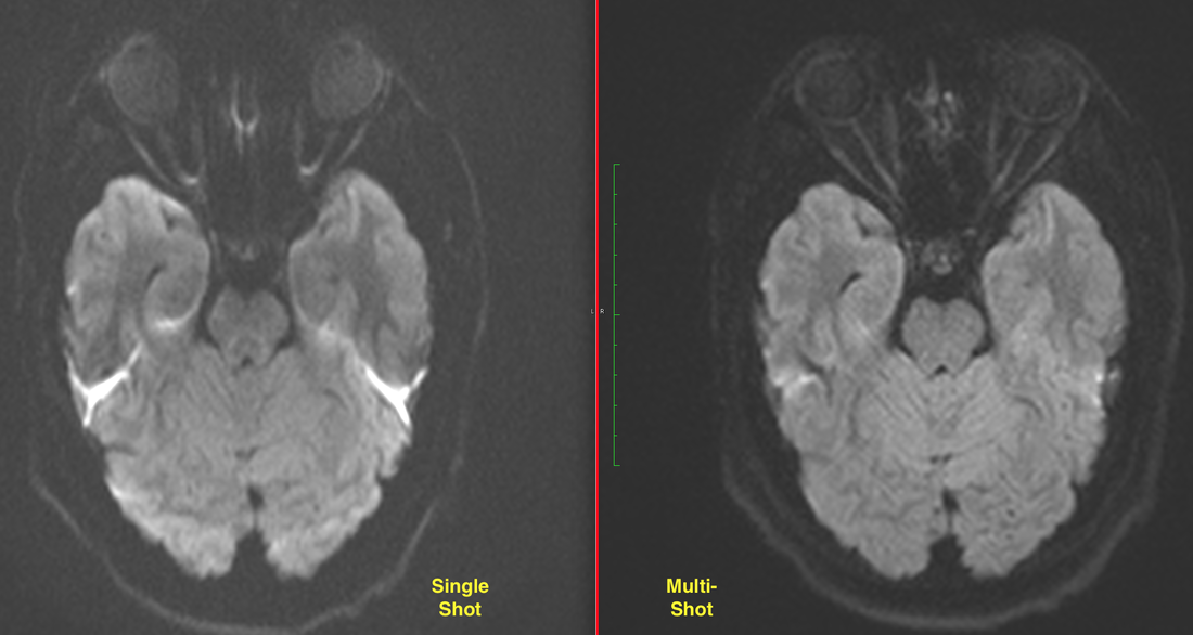
Conventional diffusion-weighted imaging (DWI) is typically performed using a single-shot echo-planar (ss-EPI) sequence. This has the advantage of speed but suffers from susceptibility artifacts/distortions, low signal-to-noise, and spatial blurring. To improve image quality (at the expense of increased imaging time) readout-segmented echo-planar diffusion methods (rs-DWI) have been developed. Siemens currently markets one such sequence under the trade name RESOLVE (REadout Segmentation Of Long Variable Echo trains).
In rs-DWI k-space sampling occurs in a small number (3-6) of "shots". Each shot has limited transversal of k-space in the readout direction, but full resolution along the phase-encode axis. Because motion artifacts between segments can create substantial artifacts, 2D navigator readout segments are acquired during the second and subsequent echoes. At k-space center, the navigator is repeatedly acquired to account for shot-dependent nonlinear phase differences that arise from non-rigid motion of the head or other imaged body part. As shown in the example below, improved signal-to-noise, spatial resolution, and decreased susceptibility artifacts are noted using this strategy
PROPELLER/BLADE diffusion methods may also be used to reduce susceptibility artifacts and improve spatial resolution compared to ss-EPI methods. These employ fast (turbo) spin echoes and sample k-space in a radial fashion. Image quality is significantly improved and motion artifacts reduced. These sequences may be superior to rs-DWI-EPI methods where susceptibility artifacts are most problematic, such as in evaluating temporal bone cholesteatomas. Notwithstanding these advantages, image acquisition time may be comparatively longer compared to rs-DWI and imaging artifacts may limit acquisition to primarily the axial plane. Additionally, the use of turbo spin echoes increases specific absorption rate (SAR) and produces more tissue heating compared to gradient echo EPI methods.
Advanced Discussion (show/hide)»
Phase-encode segmented DWI has also been tested, where all readout k-space data are sampled at each shot, but at a limited number of phase encoding points. Such sequences can provide higher spatial resolution and imaging in all orientations. However, this sampling scheme cannot be easily combined with 2D, non-linear, navigator phase correction. Accordingly, phase-encode segmented DW images are more prone to motion-induced aliasing artifacts than readout-segmented ones.
References
Morelli JN, Saettele MR, Rangaswamy RA, et al. Echo planar diffusion-weighted imaging: possibilities and considerations with 12- and 32-channel head coils. J Clin Imaging Sci 2012;2:31.
Pipe JG, Farthing VG, Forbes KP. Multishot diffusion-weighted FSE using PROPELLER MRI. Magn Reson Med 2002; 47:42-52.
Porter DA, Heidemann RM. High resolution diffusion‑weighted imaging using readout‑segmented echo‑planar imaging, parallel imaging and a two‑dimensional navigator‑based reacquisition. Magn Reson Med 2009;62:468‑75.
Related Questions
What is echo-planar imaging (EPI)? Is this the same as Fast Spin Echo (FSE)?
You alluded to different trajectories for filling k-space. What are they?
How does PROPELLER reduce motion artifacts?
What is echo-planar imaging (EPI)? Is this the same as Fast Spin Echo (FSE)?
You alluded to different trajectories for filling k-space. What are they?
How does PROPELLER reduce motion artifacts?


