|
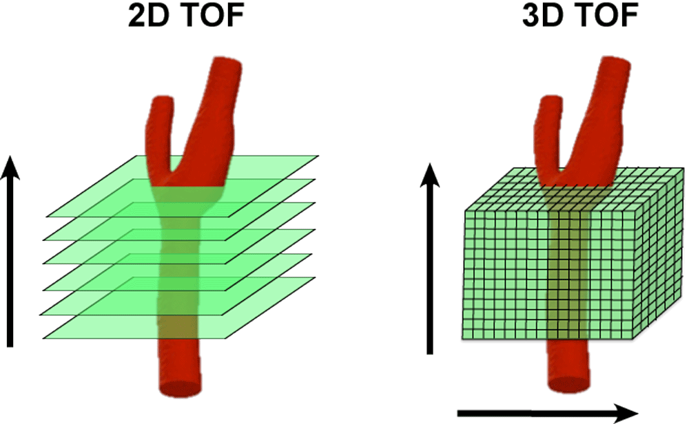
Most MRA techniques, including time-of-flight (TOF) methods, can be performed using either a 2D or 3D acquisition mode. 2D TOF is commonly used for imaging of long vascular segments running perpendicular to the plane of imaging (like the aorta or femoral arteries). The 3D mode is used for more compact anatomic regions with various flow directions (like the carotid bifurcation, circle of Willis, or renal arteries).
|
|
2D TOF MR Angiography. Multiple thin (1-2 mm-thick) slices are obtained as a stack in a plane perpendicular to the course of the imaged blood vessels. TR values are kept short (<30 ms) to suppress background tissues and accentuate flow-related enhancement. The slices in 2D TOF MRA are acquired sequentially; complete data for each slice is collected before moving on to the next slice. A traveling saturation band about 4 cm wide is generally placed on the upstream side of each slice to suppress contaminating signal from venous inflow.
The 2D method offers short imaging times with excellent sensitivity to slow flow. Imaging of long vascular segments is possible (such as the entire aorta or lower extremity arteries) by simply increasing the number of slices. Disadvantages of 2D imaging include insensitivity to in-plane flow and patient motion artifacts that may create misregistration. |
|
3D TOF MR Angiography. The 3D TOF mode is used where the imaged anatomy encompasses a relatively small area and vessels run in various orientations. 3D methods offer high spatial resolution and high signal-to-noise. Nearly isotropic (square) voxels may be obtained, allowing reformation in any direction.
Disadvantages of 3D TOF MRA include relative insensitivity to slow flow and saturation effects that limit maximum slab thickness of each acquisition. Imaging times are also longer than 2D TOF techniques. Although motion artifacts affect only single slices in 2D TOF MRA, in 3D acquisitions, all slices are affected, potentially rendering the entire study uninterpretable.
|
A brief summary of the advantages and disadvantages of 2D-TOF and 3D-TOF is provided in the table below:
Advanced Discussion (show/hide)»
No supplementary material yet. Check back soon!
References
Saloner D. An introduction to MR angiography. Radiographics 1995;15:453-465. (Older review; good discussion of TOF and PC MRA)
Saloner D. An introduction to MR angiography. Radiographics 1995;15:453-465. (Older review; good discussion of TOF and PC MRA)
Related Questions
How does time-of-flight MRA work?
How does time-of-flight MRA work?


