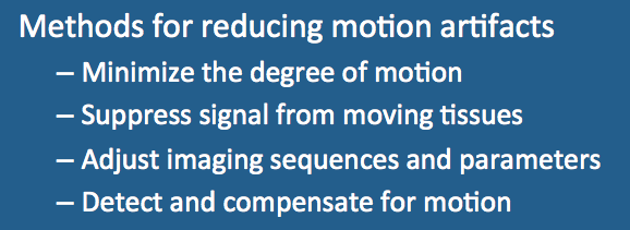
Fortunately, a large number of strategies are available for reducing or eliminating motion artifacts. These are briefly outlined below, with separate subsequent Q&A's devoted to each.
1. Minimize the degree of motion.
a. The importance of simple instruction/education of the patient to hold still while the scanner is making noise should not be underestimated. Pre-scan training and practice with breath holding may be helpful.
b. The patient should be made as comfortable in the scanner as possible, including back, leg, or head supports. Stabilization measures including the use foam pads, taping, snug wrapping with sheets, or even bite bars may be useful. Infants should be swaddled and their heads supported.
c. For uncooperative patients or those with high anxiety or pain, sedation or general anesthesia may be required.
d. Some physiologic motions such as peristalsis can be reduced with appropriate pharmacologic agents (e.g., glucagon 1 mg) given IM to prolong their action.
2. Suppress signal from moving tissues.
a. Using surface coils confined to the area of interest will minimize unwanted signals from moving tissues located farther away. A good example is the use of a spine coil array which will naturally attenuate signals from the moving anterior abdominal wall due to distance.
b. Spatial saturation pulses can null signals from unwanted moving anatomical objects.
c. Fat suppression techniques (STIR, CHESS, etc) will null high signal from subcutaneous and juxtadiaphragmatic fat stores that are often responsible for motion artifact.
d. Flow saturation pulses will suppress signals from arterial or venous blood entering a slice.
3. Adjust imaging sequences and parameters.
a. Increasing number of signals averaged (NSA, NEX) will reduce artifacts and increase signal-to-noise but at expense of increased imaging time.
b. Swapping frequency- and phase-encoding directions will shift direction of artifacts but will not reduce them.
c. Single-slice ultrafast sequences (HASTE, EPI, TrueFISP) may acquire images rapidly enough (2-5 sec) to freeze bulk motion without breath holding or any additional special techniques.
d. Radial/spiral sequences are more effective than those using Cartesian trajectories at dispersing motion artifacts throughout an image.
e. Flow compensation (FlowComp, GMN) techniques reduce artifacts from flowing blood and spinal fluid by gradient refocusing of signal.
4. Detect and compensate for motion.
a. Hardware-based gating methods for respiratory or cardiovascular motion are widely available. Respiratory expansion may be detected by use of a thoracic belt or bellows. Cardiovascular motion can be detected by EKG or peripheral pulse device. Once detected, the MR pulse sequence can be prospectively triggered to a specific time in the cardiac cycle, respiratory cycle, or both. Retrospective gating can also be performed. In this technique MR data is continuously acquired and points are reordered or discarded retroactively based on their timing within the cardiorespiratory cycle.
b. Navigator echo techniques (e.g., PACE) use additional RF-pulses to track cardiac or diaphragmatic position. Information from the navigator echo can be used prospectively to trigger data acquisition or retrospectively to adjust the location of a group of slices already acquired.
c. Self-correcting sequences (PROPELLER, BLADE) oversample the center of k-space and can detect in-plane rotation or translation. Aberrant slices can be rejected or realigned with the others through an iterative procedure.
d. Co-registration to external landmarks or a reference image can control motion artifacts over a 3-dimensional volume. Images are transformed by spatial translations, rotations, and interpolations.
Advanced Discussion (show/hide)»
No supplementary material yet. Check back soon!
References
Salem KA. Motion correction for MR imaging. Siemens Medical, 2005. (Sales brochure with a decent review of several techniques).
Zaitsev M, Maclaren J, Herbst M. Motion artifacts in MRI: a complex problem with many partial solutions. J Magn Reson Imaging 2015; 42:887-901.
Salem KA. Motion correction for MR imaging. Siemens Medical, 2005. (Sales brochure with a decent review of several techniques).
Zaitsev M, Maclaren J, Herbst M. Motion artifacts in MRI: a complex problem with many partial solutions. J Magn Reson Imaging 2015; 42:887-901.
Related Questions
Why do motion artifacts often form into discrete ghosts?
Why do motion artifacts often form into discrete ghosts?
