Radiofrequency irradiation used in MRI causes tissue heating, primarily the result of ohmic (resistive) losses due to induced electrical currents. The rate of RF energy deposition (power) absorbed per mass of tissue is called the specific absorption rate (SAR), typically reported in units of watts per kilogram (W/kg). Various regulatory organizations, like the International Electrotechnical Commission (IEC) and US Food and Drug Administration (FDA) have established safe limits for SAR deposition and temperature changes in patients undergoing MRI.
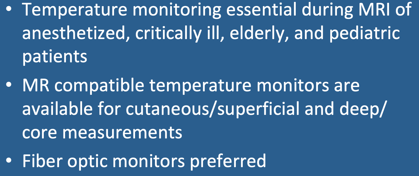
Patients at risk for thermoregulatory disturbances include infants, pregnant women, the elderly, obese, diabetics, febrile patients, anesthetized patients, and those with cardiac decompensation. Core body temperature (normal 36ºC − 38ºC) is regulated by the hypothalamus, but can disturbed by certain medications — beta-blockers, diuretics, calcium-channel blockers, amphetamines, anesthetic agents, and sedatives. For such patients, as well as any unable to effectively communicate, monitoring body temperature during MRI is required.
Patients at risk for thermoregulatory disturbances include infants, pregnant women, the elderly, obese, diabetics, febrile patients, anesthetized patients, and those with cardiac decompensation. Core body temperature (normal 36ºC − 38ºC) is regulated by the hypothalamus, but can disturbed by certain medications — beta-blockers, diuretics, calcium-channel blockers, amphetamines, anesthetic agents, and sedatives. For such patients, as well as any unable to effectively communicate, monitoring body temperature during MRI is required.
A wide range of potential sites are available for temperature measurement: skin, mouth, axilla, groin esophagus, rectum, pulmonary artery, and bladder. Superficial sites (skin, axilla, groin) are most commonly used due to their accessibility, but are affected by perspiration, ambient air circulation, humidity, use of blankets, and peripheral vasoconstriction. Deep sites (esophagus, bladder, rectum) more accurately reflect core body temperature but are more cumbersome to use and set up.
|
The simplest (and cheapest) skin thermometers are non-magnetic with a liquid crystal displays. They can be stuck on the forehead or other easily visible site and may be suitable for relatively brief MR exams to provide a modicum of reassurance concerning body temperature in otherwise non-critical patients. Thermistor and thermocouple-based sensors are subject to drift and may cause RF interference, so should be avoided in the MR environment. The current best MRI-compatible temperature probes utilize a fiber optic cable with either a fluorotopic or gallium-arsenide (GaAs) detector. Fiber optic probes contain no metal can be used for either superficial or deep temperature measurements. Rectal temperature fiberoptic probes may be acceptable for relatively stable patients undergoing 1 hour or less of MRI. For patients undergoing general anesthesia a fiber optic probe placed in the distal third of the espohagus is recommended.
|
Advanced Discussion (show/hide)»
No supplementary material yet. Check back soon!
References
Adair ER, Black DR. Thermoregulatory responses to RF energy absorption. Bioelectromagnetics 2003; 24(S6):S17-S38. [DOI Link]
Foster KR, Glaser R. Thermal mechanisms of interaction of radio frequency energy with biological systems with relevance to exposure guidelines. Health Phys 2007; 92:609-620. [DOI link]
Schena E, Tosi D, Saccomandi P, et al. Fiber optic sensors for temperature monitoring during thermal treatments: an overview. Sensors 2016; 16:1144. [DOI LINK]


