Images of the beating heart are not only aesthetically pleasing, but are critically important for the diagnosis of cardiac function. As such, multiplanar cine studies are an essential component of nearly every cardiac MR examination.
|
The base pulse sequence is a bright-blood technique, typically a balanced steady-state free precession (SSFP) gradient echo method such as TrueFISP. As described in a prior Q&A, TrueFISP generates high intravascular signal relative to other tissues due to the intrinsically high T2/T1 ratio of blood. The structure of this sequence allows very short TR and TE values to be used, and hence multiple lines of k-space (echoes) can be acquired during a single heart beat.
|
TrueFISP cine heart study
|
|
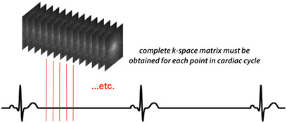
Cine studies are typically obtained by repeatedly imaging the heart at a single slice location throughout the cardiac cycle. Between 10 and 30 cardiac phases are usually sampled. To avoid confusion with other uses of the term "phase" in MRI, these individual cardiac images are often referred to as "frames" or "segments".
|
To fully evaluate the entire heart, separate cine image sets at various locations must be obtained. For example, a standard cine study might include three sets of short-axis images (base, mid-ventricle, and apex), three sets of long-axis images (2-, 3-, and 4-chamber), plus valvular and outflow tract images as needed.
To maintain SSFP conditions for bright-blood imaging, the RF-pulses must run continuously. Retrospective EKG-gating is performed so that imaging data can be assigned to the appropriate phase of the cardiac cycle.
|
Cine sequences use very short TE's (1-2 ms) permitting multiple lines of k-space to be acquired within each frame during a single heart beat (R-R interval). This value is an operator-selectable parameter known as views per frame (vps) or lines per segment (lps).
Because only a limited number of views per frame are possible, data must be collected over many cardiac cycles. Nevertheless, a complete set of cine images for a given slice location can usually be collected in 5-10 seconds, well within the single breath hold capabilities of most patients. |
Advanced Discussion (show/hide)»
No supplementary material yet. Check back soon!
References
Atkinson DJ, Edelman RR. Cineangiography of the heart in a single breath hold with a segmented TurboFLASH sequence. Radiology 1991; 178:357-360. (In the 1990s and early 2000s, cine studies were performed using a spoiled GRE sequence as in the method described here).
Carr JC, Simonetti O, Bundy J, et al. Cine MR angiography of the heart with segmented true fast imaging with steady-state precession. Radiology 2001; 219:828-834. (First full description of a TrueFISP-based cine sequence, showing its superiority over segmented TurboFLASH methods).
Related Questions
What is True FISP, and why is it "truer" than regular FISP?
What is True FISP, and why is it "truer" than regular FISP?


