|
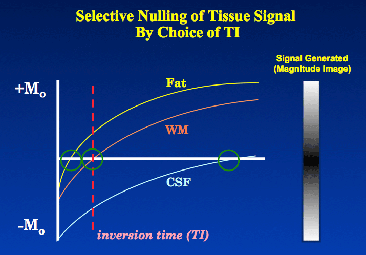
The usual Inversion Recovery sequence begins with a 180°-inverting pulse, which reverses the longitudinal magnetization (Mz) for all tissues. During the inversion time (TI) interval, these Mz's regrow via T1-relaxation seeking to restore their equilibrium alignment in the +z-direction. A spin echo (or more commonly a fast spin echo) sequence then generates a signal based on the longitudinal magnetization of each tissue at time TI.
|
If a tissue happens to have longitudinal magnetization (Mz) close to zero at the end of the TI interval, that tissue will produce little or no signal and make no significant contribution to the magnitude-reconstructed IR image. The tissue is said to be "nulled" or "suppressed". The value of TI where this happens is known as the "null point" or "zero-crossing point" and denoted TInull.
TInull depends on the ratio TR/T1 and whether the signal is generated by a conventional spin echo (CSE) or Fast/Turbo spin echo (FSE) mechanism. Two equations can be derived
TInull depends on the ratio TR/T1 and whether the signal is generated by a conventional spin echo (CSE) or Fast/Turbo spin echo (FSE) mechanism. Two equations can be derived
Note that when TR>>T1, the equations both reduce to TInull = T1 • (ln 2) ≈ 0.69 x T1.
At 1.5T and assuming a very long TR (≥ 4000 ms), TInull values for fat, white matter, and gray matter are approximately 180, 400, and 650 ms respectively. Because tissue T1 increases with field strength, these values must be adjusted upward if performing imaging at 3.0T or higher.
The T1 value of CSF and other pure liquids is extremely long (on the order of 3000-4000 ms), so usually the simplifying condition TR>>T1 is not met. Additionally since most modern clinical IR imaging uses FSE signal generation with as many as 16-32 echoes, the time of the last echo (TElast) also cannot not be ignored. Thus TInull for CSF is not a single number but depends on the precise TR and TElast values chosen.
Advanced Discussion (show/hide)»
No supplementary material yet. Check back soon!
References
Bernstein MA, King KF, Zhou XJ. Handbook of MRI pulse sequences. Chicago: Academic Press, 2004; pp. 631-5.
Bernstein MA, King KF, Zhou XJ. Handbook of MRI pulse sequences. Chicago: Academic Press, 2004; pp. 631-5.
Related Questions
Why use IR? What are the advantages?
Why use IR? What are the advantages?


