|
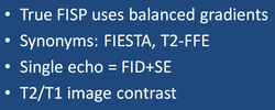
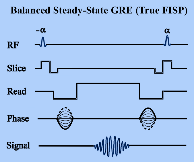
True FISP is the Siemens trade name for a steady-state coherent sequence in which balanced gradients are used along all three axes. "Balanced" means that the net gradient-induced dephasing over a TR interval is zero. Conversely, some of the gradients in FISP and PSIF are unbalanced, allowing recording of separate FID and echo components of the steady-state free precession signal. In True FISP, however, the balanced gradients refocus both components at the exact center of the TR interval as a single echo. Other vendors have similar sequences. GE calls theirs FIESTA (Fast Imaging Employing Steady-state Acquisition) and Philips calls theirs Balanced-FFE (Fast Field Echo) respectively.
|
|
Although the principles underlying echo formation in True FISP have long been known, widespread clinical implementation has been slow due to stringent technical requirements. True FISP sequences demand a very high level of magnetic field homogeneity and control over gradient switching and shaping. The refocusing mechanism fails if intravoxel dephasing exceeds ±180º manifest by band-like artifacts. During the last decade modern scanners have overcome these limitations making True FISP a viable and useful sequence on most mid- and high-field systems.
|
The signal intensity (SBal) for the balanced (True FISP) sequence is given by
When the echo is recorded close to the middle of the interval (TE ≈ TR/2, as is usually the case), the final term e−TE/T2 depends on T2, not T2*. Thus True FISP sequences behave more like spin echo than gradient echo sequences in that they do not have T2*-dependence. Also, since TR is nearly always much, much shorter than T1 or T2, the exponential terms containing TR can be disregarded and the equation simplified to
|
This is an interesting result. The signal intensity is seen to be directly related to the ratio 1 ÷ (T1/T2) = T2/T1. For most solid tissues, the T2/T1 ratio is small. The exceptions are fat (0.30) and fluids (0.70). Thus True FISP sequences tend to highlight fluids such as CSF and blood, making them ideal for cardiovascular MRI, MR cisternography/myelography, MR urography, and MR enterography.
|
|
For most True FISP applications where bright T2/T1 signal is desired, TR is chosen to be a short as possible, TE = TR/2, and flip angle (α) is set in a moderate to high range (50°-80°). Low flip angles provide spin density weighted contrast but are seldom used. Long TR values are likewise usually avoided.
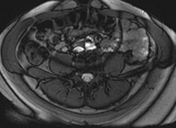
Left: Cardiac True FISP image with TR/TE/α = 7.0/3.5/70° |
Advanced Discussion (show/hide)»
No supplementary material yet. Check back soon!
References
Bieri O, Scheffler K. Fundamentals of balanced steady state free precession MRI. J Magn Reson Imaging 2013;38:2-11.
Huang T-Y, Huang I-J, Chen C-Y, et al. Are TrueFISP images T2/T1-weighted? Magn Reson Med 2002; 48:684-688. (Yes, but not so much during the initial period during transition to a full steady-state).
Scheffler K, Hennig J. Is TrueFISP a gradient-echo or a spin-echo sequence? Magn Reson Med 2003; 49:395-7. (It's more like SE than GRE, as long as the echo is centered).


