|
When appropriate imaging parameters are used, GRE sequences generate a strong signal from moving spins, highlighting flowing blood or CSF. This property has made GRE-based sequences the backbone of most MR angiographic techniques. GRE sequences accentuate signals from flowing fluids for three principal reasons.
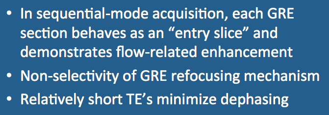
First, when sequential mode acquisition (rather than interleaved multi-slice mode) is employed, each section behaves like an "entry slice" and demonstrates flow-related (inflow) enhancement. This statement is true for any flip angle, as long as the sequence is gradient- or RF-spoiled or the TR value is long enough to minimize steady-state effects.
|
For steady-state GRE sequences (e.g., GRASS/FISP), flow sensitivity can be increased by using moderate-to-large flip angles. Large flip angles accentuate the signal from flowing blood or CSF for two reasons: (1) they cause more saturation of stationary tissue, and when unsaturated spins flow in, a greater contrast between moving and static tissue can be appreciated; and (2) large flip angles produce signal intensity differences proportional to T2/T1, a ratio that is higher in CSF, blood, and other fluids than for solid tissues.
The second factor increasing the flow sensitivity of GRE imaging concerns the nonselectivity of the GRE refocusing mechanism. In routine SE imaging, at least part of the reason for flow-related signal loss is that spins move out of the section between the 90°- and 180°-(refocusing)-pulses. In GRE imaging the refocusing is done by means of a gradient reversal that is not slice-selective. This nonselective refocusing does not result in hyperintensity of incoming spins, however; it merely prevents them from experiencing TOF signal losses.
Finally, relatively short echo times (TE's) are often used in conjunction with GRE techniques to minimize T2*-dephasing. Because these short echo times also minimize flow-related signal losses, they accentuate the appearance of the flowing fluid.
Advanced Discussion (show/hide)»
No supplementary material yet. Check back soon.
References
Atlas SW, Mark AS, Fram EK, Grossman RI. Vascular intracranial lesions: applications of gradient-echo MR imaging. Radiology 1988; 169:455-461.
Atlas SW, Mark AS, Fram EK, Grossman RI. Vascular intracranial lesions: applications of gradient-echo MR imaging. Radiology 1988; 169:455-461.
Related Questions
How can you predict whether a certain vessel will be bright or dark on an MR image?
How can you predict whether a certain vessel will be bright or dark on an MR image?

