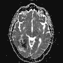
T2-blackout is the opposite of the T2-shine-through phenomenon, described more completely in a prior Q&A. In T2-blackout, lesions with very short T2 (or T2*) values reduce signal intensity in the DW image, potentially masking or destroying its diffusion sensitivity. In extreme cases even the ADC map calculation will be affected and unreliable.
An example of T2-blackout is shown below for a subacute hematoma. The T2*-weighted b0 image has extremely low signal due to the paramagnetic effects of intracellular deoxyhemoglobin. The trace DW image also has a dark center, but this is primarily due to blackout from the T2 (T2*) effects. The ADC map shows a scattered mix of bright and dark pixels within the hematoma, demonstrating the computational hazards of estimating ADC values in such situations.
An example of T2-blackout is shown below for a subacute hematoma. The T2*-weighted b0 image has extremely low signal due to the paramagnetic effects of intracellular deoxyhemoglobin. The trace DW image also has a dark center, but this is primarily due to blackout from the T2 (T2*) effects. The ADC map shows a scattered mix of bright and dark pixels within the hematoma, demonstrating the computational hazards of estimating ADC values in such situations.
The reason both T2-shine-through and T2-blackout occur relate to the nature of the DW (trace) image. By definition the DW image is diffusion-weighted, not a map of diffusion. The DW image is strongly dependent on diffusion, but also reflects underlying T2, spin-density, and T1 effects. When the intrinsic T2 of a lesion is long, T2 effects may spill over into the DW image, making it appear bright and thus mimicking restricted diffusion ("T2-shine-through"). When the T2 (or T2*) is very short, the opposite occurs, with resulting decrease in signal intensity on the DW image ("T2-blackout").
Advanced Discussion (show/hide)»
Note in the middle DW image above, a bright halo is also present around the edge of the hematoma that is not the same as the ring of vasogenic edema surrounding the entire lesion. This halo is a susceptibility artifact due to the paramagnetic effects of deoxyhemoglobin. Susceptibility artifacts will be discussed in a later Q&A.
References
Hiwatashi A, Kinoshita T, Moritani T et-al. Hypointensity on diffusion-weighted MRI of the brain related to T2 shortening and susceptibility effects. AJR Am J Roentgenol 2003; 181:1705-9.
Maldjian JA, Listerud J, Moonis G, Siddiqi F. Computing diffusion rates in T2-dark hematomas and areas of low T2 signal. AJNR Am J Neuroradiol 2001;22:112–128.
Silvera S, Oppenheim C, Touzé E, et al. Spontaneous intracerebral hematoma on diffusion-weighted images: influence of T2-shine-through and T2-blackout effects. AJNR Am J Neuroradiol 2005; 26:236-242.


