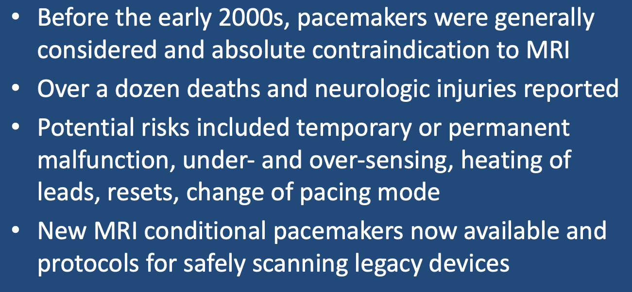
Until the last two decades, the presence of a cardiac pacemaker was considered an absolute contraindication for MR imaging. The traditional 5 Gauss safety line (beyond which the general unscreened public must not pass) is still in use, having been based upon the approximate field at which the reed switches in older pacemakers may be affected. The reasons traditionally cited in support of excluding pacemaker patients from MRI include:
- Over a dozen documented deaths and non-fatal but serious injuries (such as hypoxic-ischemic encephalopathy from cardiac arrest) have been reported in pacemaker patients inadvertently subjected to MR scanning. The actual worldwide number of similar but unreported events is likely to be much higher.
- Temporary pacemaker malfunction during the scan leading to cardiac arrhythmias or impaired cardiac output can occur due to inhibition of pacing output, over- or under-sensing, unpredictable opening/closure of the reed switch, alteration of programming, overly rapid pacing, and device power-on reset.
- Permanent electromagnetic field damage to pacemaker electronics or battery necessitating pacemaker replacement may occur.
- RF-induced heating at the electrode tip via the antenna effect (see separate Q&A) has been demonstrated experimentally and may be manifest by increased lead resistance/capture threshold.
- An additional hazard unique to internal cardiac defibrillators (ICD's) exists — false interpretation of RF-pulses as ventricular tachyarrhythmia and deliverance of cardioversion/defibrillation shocks.
As the turn of the century approached, some centers (including ours) began to perform MRI's in selected pacemaker patients for whom clinical benefit was thought to outweigh these potential risks. We did this with some trepidation and were exceedingly cautious at first, initially having cardiologist transvenous pacer in hand and a cardiac nurse ready with drugs in the event of an emergency. But over time, we learned that this heightened level of preparedness was not usually necessary and that many patients with even older pacemakers could be scanned safely with thorough pre-procedure evaluation by a cardiologist and radiologist and moderate supervision during the scan. In the mean time, the medical device industry responded by producing MR Conditional pacemakers, the first models appearing in 2007, with many more continuing to be developed.
Advanced Discussion (show/hide)»
No supplementary material yet. Check back soon!
References
Gupta SK, Ya’qoub L, Wimmer AP, et al. Safety and clinical impact of MRI in patients with non-MRI-conditional cardiac devices. Radiology: Cardiothoracic Imaging 2020; 2(5):e200086. [DOI LINK]
Luechinger R, Duru F, Zeijlemaker VA, et al. Pacemaker reed switch behavior in 0.5, 1.5, and 3.0 Tesla magneict resonance imaging units: are reed switches always closed in strong magnetic fields. Pacing Clin Electrophysiol 2002; 25:1419-23. (reed switches closed at 8G±2 and opened at 7G±2, supporting the long-held belief of the 5G line as still valid)
Mulpuru SK, Madhavan M, McLeod CJ, et al. Cardiac pacemakers: function, troubleshooting, and management. J Am Coll Cardiol 2017; 69:189-210. [DOI LINK]
Muthalaly RG, Nerlekar N, Ge Y, et al. MRI in patients with cardiac implantable electronic devices. Radiology 2018; 289:281-292. [DOI LINK]
Poh PG, Lieu C, Yeo C, et al. Cardiovascular implantable electronic devices: a review of the dangers and difficulties in MR scanning and attempts to improve safety. Insights Imaging 2017; 8:405-418. [DOI LINK]
Rajah P, Kay F, Bolen M, et al. Cardiac magnetic resonance in patients with cardiac implantable electronic devices. Challenges and solutions. J Thor Imaging 2020; 35:W1-W17. [DOI LINK]
Roguin A, Schwitter J, Vahlhaus C, et al. Magnetic resonance imaging in individuals with cardiovascular implantable electronic devices. Europace 2008; 10:336-346. [DOI LINK]
Shinbane JS, Colletti PM, Sherlock FG. Magnetic resonance imaging in patients with cardiac pacemakers: era of “MR Conditional” designs. J Cardiovasc Magn Reson 2011;13:63. [DIRECT LINK]
Vigen KK, Reeder SB, Hood MN, et al. Recommendations for imaging patients with cardiac implantable electronic devices (CIEDs). J Magn Reson Imaging 2020. [DOI LINK]
Related Questions
What are the risks of passive vs active implants in MRI?
I'm not a cardiologist. Can you explain some of the pacemaker types and terminology relevant to us working in MRI?
What are the risks of passive vs active implants in MRI?
I'm not a cardiologist. Can you explain some of the pacemaker types and terminology relevant to us working in MRI?
