|
Moran analyzed the phase effects on stationary and moving spins subjected to a pair of bipolar gradients as illustrated in the diagram right. A stationary spin subjected to such a gradient pair will experience no net phase shift, but a moving spin will have a net phase shift proportional to its velocity. Two spins flowing at the same speed but in opposite directions will have equal but opposite phase shifts. By measuring changes in phase, therefore, velocity can be computed.
|
Advanced Discussion (show/hide)»
In practice using simple bipolar gradients to measure flow velocities is not possible because magnetic field inhomogeneities create additional phase shifts that are indistinguishable from motion. To correct for these unwanted phase shifts, a subtraction technique is used to remove the background phase accrual. Typically, this involves toggling the bipolar gradients: (+/−) followed by (−/+) and subtracting the paired data.
To fully describe motion in all directions and create an MR angiogram, the bipolar subtraction method would require up to six separate data acquisitions (2 toggled pairs applied along 3 axes). More efficient methods using only four acquisitions have been developed and are used on most commercial systems. These involve either using a common 3-axis velocity-compensated reference image for subtraction or a Hadamard encoding scheme toggling bipolar gradients along two axes simultaneously.
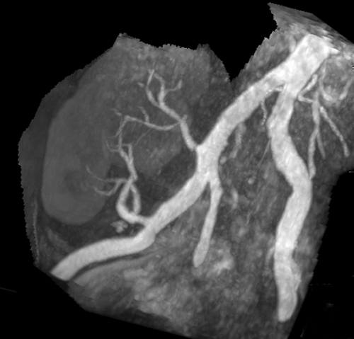
Even with these efficiencies, the general use of 3D-PC as an angiographic method has greatly declined since the 1990s, mainly because of its long imaging times (1.5x−2.0x) that of equivalent 3D-TOF-MRA sequences. PC is still a relatively common method to perform MRA of renal arteries, renal transplants, and mesenteric vessels. Intracranially PC MRA is still used in relatively niche situations such as to evaluate AVM's and for aneurysm workups in the presence of hemorrhage (the T1 shine-through effect of blood products on TOF MRA does not occur with PC MRA).
The widespread use of parallel imaging techniques in conjunction with PC-MRA significantly reduces its imaging time penalty. I wouldn't be surprised to see PC-MRA being used more in the future.
Bryant DJ, Payne JA, Firmin DN, et al. Measurement of flow with NMR imaging using a gradient pulse and phase difference technique. J Comput Assist Tomogr 1984;8:588-93.
Dyverfeldt P, Bissell M, Barker AJ, et al. 4D flow cardiovascular magnetic resonance consensus statement. J Cardiovasc Magn Reson 2015; 17:72.
Firmin DN, Nayler GL, Klipstein RH, et al. In vivo validation of MR velocity imaging. J Comput Assist Tomogr 1987;11:751-6.
Lotz J, Meier C, Leppert A, Galanski M. Cardiovascular flow measurement with phase-contrast MR imaging: basic facts and implementation. Radiographics 2002; 22:651-671.
Markl M, Frydrychowicz A, Kozerke S, et al. 4D Flow MRI. J Magn Reson Imaging 2012; 36:1015-1036.
Moran PR. A flow velocity zeugmatographic interlace for NMR imaging in humans. Magn Reson Imaging 1982; 1:197-203.
Moran PR, Moran RA, Karstaedt N. Verification and evaluation of internal flow and motion. True magnetic resonance imaging by the phase gradient modulation method. Radiology 1985;154:433-441.
Nayak KS, Nielsen J-F, Bernstein MA, et al. Cardiovascular magnetic resonance phase contrast imaging. J Cardiovasc Magne Reson 2015; 17:71.
Nayler GL, Firmin DN, Longmore DB. Blood flow imaging by cine magnetic resonance. J Comput Assist Tomogr 1986;10:715-22.
Stankovic Z, Allen BD, Garcia J, et al. 4D flow imaging with MRI. Cardiovasc Diagn Ther 2014; 4:173-192.
Wildermuth S, Debatin JF, Huisman TAGM, et al. 3D phase contrast EPI MR angiography of the carotid arteries. J Comput Assist Tomogr 1995; 19:871-878.
What are spin-phase effects?


