|
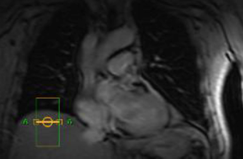
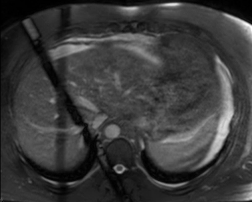
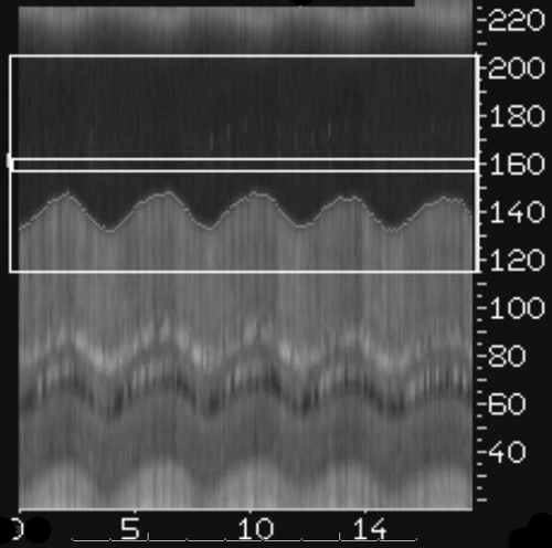
The echo signal returned by the navigator is reconstructed in the readout (motion) direction and displayed as lines of data along the beam direction. This technique is a form of line-scan imaging. It is also known as M-mode display since is equivalent to that used for viewing valve motion in M-mode echocardiography.
Automated software detects the diaphragmatic peaks and troughs of motion. The MR technologist can further define at which positions data acquisition is permitted of excluded, usually within a certain tolerance, typically 3-6 mm.
|
Advanced Discussion (show/hide)»
Additional Comments about Navigators:
One parallel imaging coil can be assigned to detect the navigator, or the entire body coil can be used.
Navigators may be leading, trailing, or both. The trailing navigator can be used to determine whether the diaphragm has returned to the same position before and after data acquisition. If the difference in position for the leading and trailing navigators exceeds a predefined threshold, the corresponding data can be discarded and remeasured.
Slice Tracking (also known as slice following) moves the scan volume to lie at approximately the same location with respect to patient anatomy for each acquisition. With tracking the position of the imaging stack is dynamically adjusted to the actual position of the anatomy of interest. A "leading navigator" is required. Both gating and tracking can be performed simultaneously.
Navigator gating levels may drift during acquisition. Automatic detection schemes are available on most scanners to correct for this by continuously monitor and update the expiration level during the scan.
Multiple navigator beams are possible on some scanners.
Two-dimensional navigator methods (e.g., Siemens' 2D PACE and Philips' MotionTrak) offer some advantages over line-scan methods. 2D methods excite a circular column of spins using excitation with a spiral k-space trajectory. This creates less extensive saturation artifacts, especially with small flip angles.
2D spiral RF-navigators may be modified by changing flip angle and number of cycles. Fewer cycles leads to a shorter pulse duration and fewer off-resonance problems but with more aliasing ring artifact near the beam. The shape of the 2D-RF pulse is typically either block or jinc, the latter having a superior excitation profile.
Ehman RL, Felmlee JP. Adaptive technique for high-definition MR imaging of moving structures. Radiology 1989; 173:255-263.
Welch EB, Manduc A, Grimm RC et al. Spherical navigator echoes for full 3D rigid body motion measurements in MRI. Magn Reson Med 2002; 47:32-41.
How can navigators track heart position if placed on the diaphragm?


