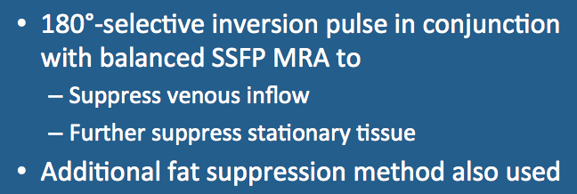
The relative high signal of arterial blood on balanced steady-state free precession (b-SSFP) sequences can be further accentuated by exploiting inflow-related enhancement similar to that used in time-of-flight MRA.
Major vendors offer such sequences under a variety of trade names. Siemens calls theirs NATIVE TrueFISP ("Non-contrast MRA of ArTerIes and VEins using Sampling Perfection with Application optimized Contrasts using different flip angle Evolution and True Fast Imaging with Steady-state Precession.") GE's version is called Inhance Inflow IR (IFIR), while Philips uses B-TRANCE ("Balanced TRiggered Angiography Non-Contrast Enhanced). Hitachi calls their method VASC-ASL ("Veins and Arteries Sans Contrast - Arterial Spin Labeling") and Cannon Time-SLIP ("Time Spatial Labeling Inversion Pulse)".
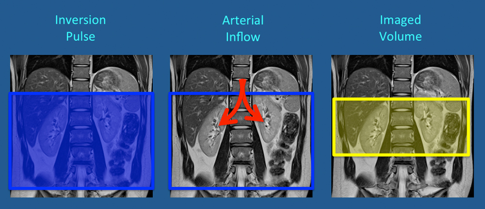
A simple workflow example illustrating the technique for renal MRA is shown below. The area of interest is first saturated with one or more 180°-RF pulses. These saturation pulses invert signal from background tissue as well as venous blood. "Fresh" (unsaturated/fully magnetized) arterial blood next flows into the slab. After an appropriate inversion time (TI) that nulls signal from background tissue, MRA signal generation is initiated using a rapid 3D b-SSFP sequence.
|
For imaging the upper abdomen or chest, cardiac gating with respiratory triggering or navigator echoes is commonly employed to reduce motion artifacts. An optional chemical shift selective fat suppression pulse (e.g. SPIR) is also frequently applied immediately before readout. Alternatively, water excitation or a Dixon method may be used.
|
 Inflow-enhanced b-SSFP using two
inversion slabs at 1200 and 400 msec
The usual imaging parameters at 1.5T have TI's in the 1000-1300 msec range. Obviously a single TI choice cannot null all tissues, but this is a compromise value that suppresses muscle and venous blood while allowing adequate time for fresh arterial blood inflow from outside the slab. For patients in cardiac failure or very slow vascular inflow due to stenoses, sometimes the TI value must be extended over more than one cardiac cycle to the range of 2000 or more.
In some cases additional obliquely oriented saturation bands may be placed over the kidneys as well. To further suppress signal from the inferior vena cava, a third inversion band is sometimes applied inferior to the larger first (blue) band at an intermediate TI value (e.g., 400-800 msec). Many variations are possible.
In some cases additional obliquely oriented saturation bands may be placed over the kidneys as well. To further suppress signal from the inferior vena cava, a third inversion band is sometimes applied inferior to the larger first (blue) band at an intermediate TI value (e.g., 400-800 msec). Many variations are possible.
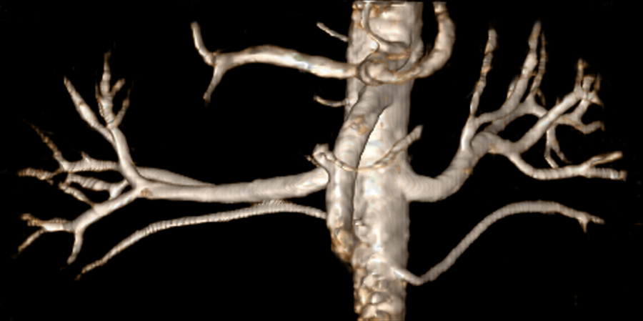
The typical balanced-SSFP sequence is performed in 3D mode, using short TR/TE values and high flip angles (e.g., TR/TE/α = 7/3/80º). A rectangular field-of-view is often used with phase oversampling to prevent wrap-around artifacts. Examples are shown below.
Like many noncontrast abdominal MRA techniques, IR-prepped balanced SSFP works well in relatively healthy patients with normal vascular flows who can hold still and maintain a regular respiratory pattern, but produces suboptimal images in sick or uncooperative patients.
Advanced Discussion (show/hide)»
Inflow-enhanced balanced SSFP MRA techniques are sometimes classified as a type of arterial spin labeling (ASL) method. I do not like this classification because the fresh blood flowing into the saturated slab has not been specifically labelled by an RF-pulse as would occur in a true ASL sequence. The inflowing blood gives off a high signal because it remains fully magnetized (unlike the saturated blood and background tissue within the imaging volume). So to me this is more of an IR-based saturation technique more similar to TOF MRA than a spin-labeling technique.
References
Katoh M, Buecker A, Stuber M, et al. Free-breathing renal MR angiography with steady-state free-precession (SSFP) and slab-selective spin inversion: initial results. Kidney Internat 2004; 66:1272-1278.
Rick M, Kaarmann N, Weale P, Schmitt P. How I do it: Non contrast-enhanced MR angiography (syngo NATIVE). MAGNETOM Flash 2009; 3:18-23.


