Magnetization transfer imaging (MTI) is a technique by which radiofrequency (RF) energy is applied exclusively to the bound pool using specially designed MT pulse(s). Some of this energy is then transferred to the free water pool primarily via dipole-dipole interactions. Depending on the degree of coupling between these pools, the free water pool becomes partially saturated. If the free water pool is subsequently imaged using routine RF pulses and gradients, its signal will be reduced secondary to the MT effect.
The magnitude of this MT effect can be quantified by obtaining two sets of images (one with an MT pulse and one without it) and then digitally subtracting them. The magnetization transfer ratio (MTR) for a given voxel may then be defined and computed as:
|
Notwithstanding these limitations, several useful clinical applications of MTC have been demonstrated. I personally find it helpful to think of MT pulses as "macromolecule sat pulses" which selectively suppress tissues with significant water-macromolecule interactions.
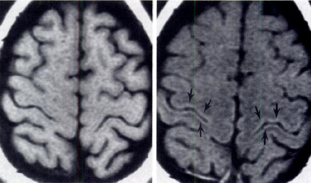
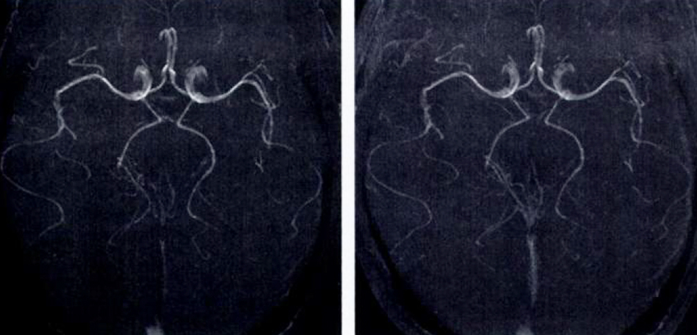
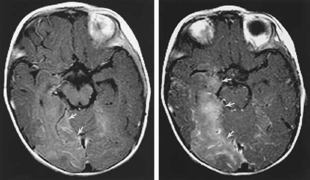
This suppression of background tissue by MT pulses makes the technique especially useful as an adjunct to MR angiography (MRA). Small vessel detail and overall image contrast are significantly improved in time of flight (TOF) MRA when MT pulses are employed. A second potential use of MT pulses is in conjunction with contrast-enhanced T1-weighted images. Because gadolinium (Gd) enhancement is caused by a water-Gd ion interaction (not macromolecular cross-relaxation), MT pulses suppress the signal from background tissues and render Gd-enhanced areas more conspicuous. A third potential use of MT pulses is in conjunction with T2-weighted images when early demyelination or protein destruction may be detected before its appearance on routine images. In this application, diseased tissues with altered protein-water interactions are less suppressed by MT pulses, rendering them more conspicuous on T2-weighted images. |
|
Advanced Discussion (show/hide)»
Additional notes on Magnetization Transfer
In the older NMR literature the term spin diffusion is sometimes used to refer to the same concept as magnetization transfer. This terminology is confusing because it is not the same as molecular diffusion which is a separate phenomenon.
The relative contributions of dipole-dipole interactions and chemical exchange to the magnetization transfer phenomenon are still being worked out. The current best evidence is that chemical exchange plays no more than a minor role in this process for most biological systems.
When an off-frequency MT pulse is applied to the system to saturate the bound pool, there is some obligatory spin-lock relaxation effects on the free water pool that contributes to its partial saturation. See the Dyke Award-winning paper by John Ulmer in the reference list for a more complete discussion of this.
Advanced techniques include MTR histograms and z-spectra. An MTR histogram is a plot of the distribution of MTRs across all pixels in an image, and shifts in such histograms have been used to follow patients with diffuse progressive brain diseases such as multiple sclerosis. An MTR z-spectrum is a plot of signal intensity vs frequency offset of the MT pulse which may change or become asymmetric in brain tumors and other disease states.
de Boer RW. Magnetization transfer contrast. Part 1: MR Physics. Philips Medical Systems MedicaMundi 1995;40:64-73.
de Boer RW. Magnetization transfer contrast. Part 2: Clinical applications. Philips Medical Systems MedicaMundi 1995;40:74-83.
Edzes HT, Samulski ET. Cross-relaxation and spin diffusion in the proton NMR of hydrated collagen. Nature 1977;265:521-523. (A famous paper first showing how the interaction between water and proteins affects relaxation times.)
Elster AD, King JC, Mathews VP, Hamilton CA. Cranial tissues: appearance at gadolinium-enhanced and nonenhanced MR imaging with magnetization transfer contrast. Radiology 1994; 190:541-546.
Elster AD, King JC, Mathews VP, Hamilton C. Improved detection of gadolinium enhancement using magnetization transfer imaging. Neuroimaging Clin N Am 1994; 4:185-192.
Mathews VP, Elster AD, King JC et al. Combined effects of magnetization transfer and gadolinium in cranial MR imaging and MR angiography. AJR Am J Roentgenol 1995; 164:169-172.
Henkelman RM, Stanisz GJ, Graham SJ. Magnetization transfer in MRI: a review. NMR Biomed 2001;14:57-64.
Ulmer JL, Mathews VP, Hamilton CA, Elster AD, Moran PR. Magnetization transfer or spin-lock? An investigation of off-resonance saturation pulse imaging with varying frequency offsets. AJNR Am J Neuroradiol 1996; 17:805-819.
Wolff SD, Balaban RS. Magnetization transfer contrast (MTC) and tissue water proton relaxation In vivo. Mag Reson Med 1989; 10: 135-144.
How does the presence of macromolecules affect T1 and T2?
What is magnetization transfer?


