|
Sternal Wires
Closure of a midline sternotomy incision using wires is the standard method used for decades. As the wires are typically made of nonferromagnetic stainless steel, titanium, or alloys, they are MR Conditional but can be scanned immediately after placement at all field strengths up to 3T. This is true even if the wires are broken. I am aware of no clinical reports of burns or discomfort of patients with sternal wires undergoing MRI, but theoretical models suggest that temperature increases of up to 10ºC may occur secondary to the antenna effect and capacitive mutual coupling between wires in certain situations. This is more likely to occur at higher field strengths and when the sutures are not simple loops, but placed in figure-of-8 or more complex configuration. Sternal Fixation Devices |
Alternatives to traditional wire closure exist. Plate and screw systems include the SternaLock® Blu (Zimmer Biomet) and Titanium Sternal Fixation System (DePuy Synthes). Other approaches include the Sternal Talon® (KLS-Martin) and an array of stainless steel and titanium-based cables and banding devices. The Sternal ZIPFIX® (DePuy Synthes) is entirely plastic and is essentially a "zip-tie" wrapped around the sternum.
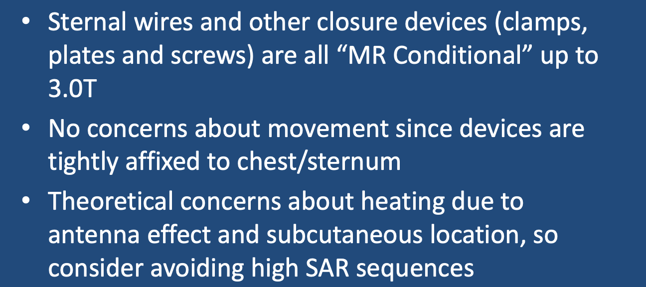
As these metallic devices are tightly adhered to the sternum, there are no concerns about displacement in the magnetic field. In that they are conductive implants with a relatively large amount of metal close to the skin surface, they are considered MR Conditional and justifiable concern exists that significant RF-heating may occur. Caution is advised to limit SAR-intensive sequences and to warn the patient to report heating or discomfort. To my knowledge no injuries serious injuries have occurred, but some patients have reported twitching of the chest muscles due to local tissue eddy currents during rapid gradient stimulation.
Pectus (Nuss) Bar
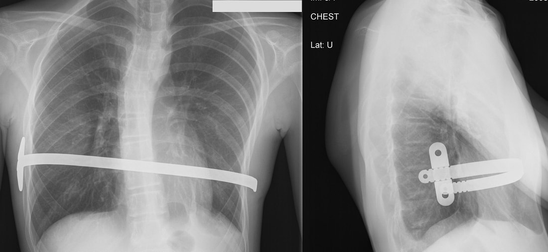
Patients with anterior thoracic wall deformities may be treated by implantation of a metallic bar across the chest anchored to the ribcage and (usually) passing under the sternum. The procedure and bar are named after their inventor, Dr. Donald Nuss, who developed this technique for pectus excavatum. To my knowledge only a single company manufactures these bars (Zimmer Biomet), which are made of titanium and rated MR Conditional up to 3T.

"MR Conditional" Pectus (Nuss) Bar. From Wikipedia under CC BY-SA 3.0, Link
Advanced Discussion (show/hide)»
A potential future device for treating pectus excavatum (now in clinical trials) is the Magnetic Mini-Mover. The system consists of a titanium-encased rare earth magnet implanted subcutaneously onto the lower sternum together with an external removable brace containing another magnet. The system is considered MR Unsafe at all field strengths.
References
Letgeb N, Loos G, Ebner F. MRI-induced tissue heating at metallic sutures (cerclages). Journal of Electromagnetic Analysis and Applications; 2013; 5:354-358.
Zheng J, Xia M, Kainz W, Chen J. Wire‐based sternal closure: MRI‐related heating at 1.5 T/64 MHz and 3 T/128 MHz based on simulation and experimental phantom study. Magn Reson Med 2020; 83:1055–1065. [DOI LINK]
Related Questions
Can one safely scan patients with total joints and other orthopedic hardware like plates and screws ?
Can one safely scan patients with total joints and other orthopedic hardware like plates and screws ?


