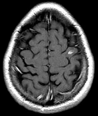
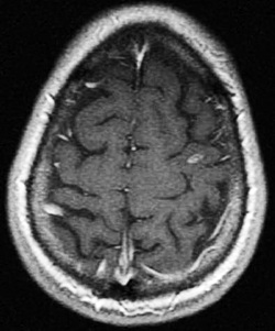
A variety of T1-weighted pulse sequences are available for postcontrast imaging. These include conventional spin echo (SE), fast (turbo) spin echo (FSE), T1-weighted fluid attenuated inversion recovery (T1-FLAIR), spoiled gradient echo (SPGR), and magnetization prepared gradient echo (MP-RAGE) methods. All provide T1-weighted contrast and are effective at showing regions of T1 shortening due to gadolinium accumulation.
Recall the important concept from a previous Q&A — that "T1-weighted" images are not "T1-maps." All T1-weighted images contain signal contributions not only from T1, but from proton density, T2, chemical shift, flow, and susceptibility effects. And each class of pulse sequences (SE, GRE, IR, etc) admix these non-contributions in different ways.
Accordingly, no single "best" gadolinium pulse sequence exists for all lesions in all locations. Considerable research has been directed to brain metastasis, however, where the conspicuity of larger lesions seems to follow the favored order T1-FLAIR > T1-SE > T1-GRE. An example is shown below.
However, T1-GRE sequences can be easily acquired in thin-section (1-2 mm) 3D mode. Hence 3D-GRE sequences may detect small metastases that might be missed on thicker 2D T1-FLAIR or 2D-SE sequences, though the latter might be superior for detecting contrast in larger lesions. In fact, for brain metastases at our institution, we use all three sequences.
For body imaging applications, the dominant considerations for optimal post-contrast pulse sequence depend on the presence of fat (which requires suppression) and motion (that must be reduced or eliminated). Again many different strategies will evolve depending on the particular region of interest or pathology considered.
Advanced Discussion (show/hide)»
No supplementary material yet. Check back soon!
References
Downs RK, Bashir MH, Ng, CK, Heidenreich JO. Quantitative contrast ratio comparison between T1 (TSE at 1.5T, FLAIR at 3T), magnetization prepared rapid gradient echo and subtraction imaging at 1.5T and 3T. Quant Imaging Med Surg 2013; 3:141-146.
Furutani K, Harada M, Mawlan, Nishitani H. Difference in enhancement between spin echo and 3-dimensional fast spoiled gradient recalled acquisition in steady state magnetic resonance imaging of brain metastasis at 3-T magnetic resonance imaging. J Comput Assist Tomogr 2008; 32:313-319.
Graves MJ. Pulse sequences for contrast-enhanced magnetic resonance imaging. Radiography 2007; 13: e20-e30.
Mirowitz SA. Intracranial lesion enhancement with gadolinium: T1-weighted spin-echo versus three-dimensional Fourier transform gradient-echo MR imaging. Radiology 1992; 185:529-534.
Related Questions
I'm confused. Isn't a spoiled-GRE sequence T1-weighted by definition?
What is T1-FLAIR?
I'm confused. Isn't a spoiled-GRE sequence T1-weighted by definition?
What is T1-FLAIR?


