|
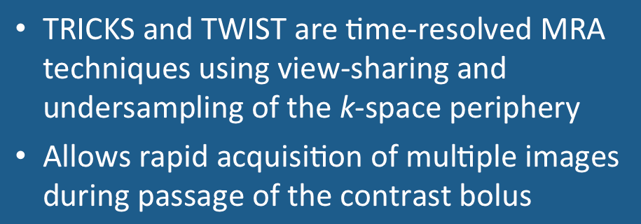
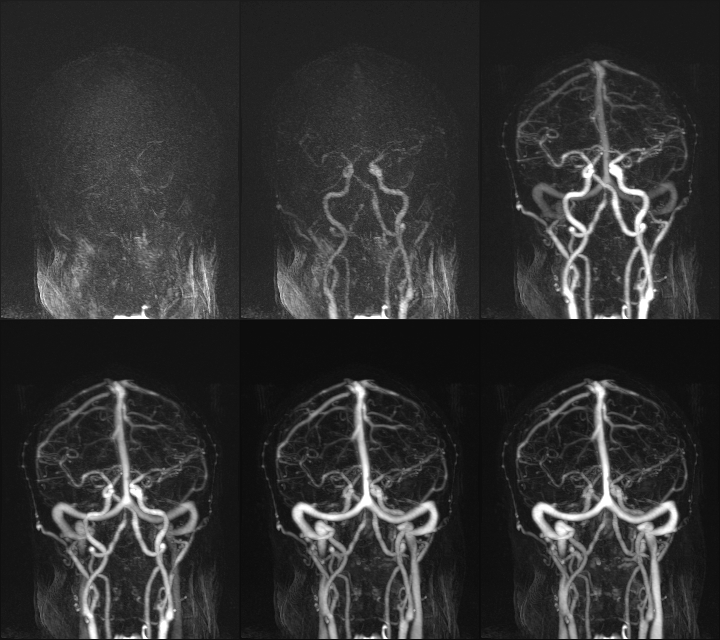
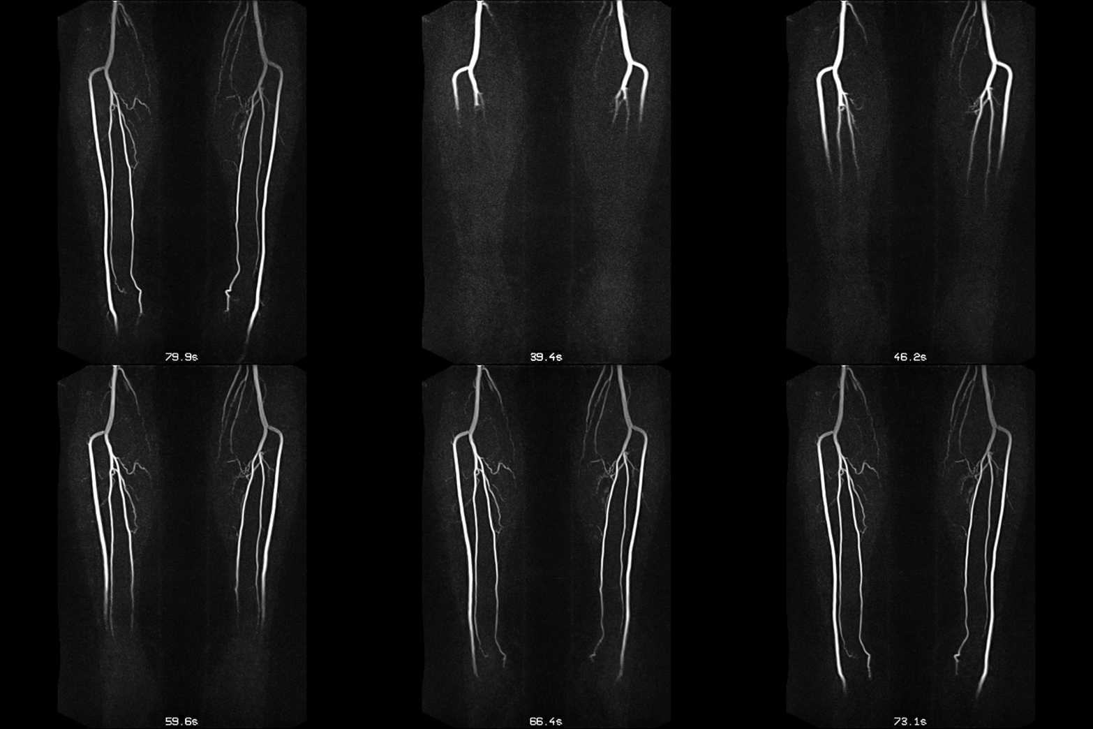
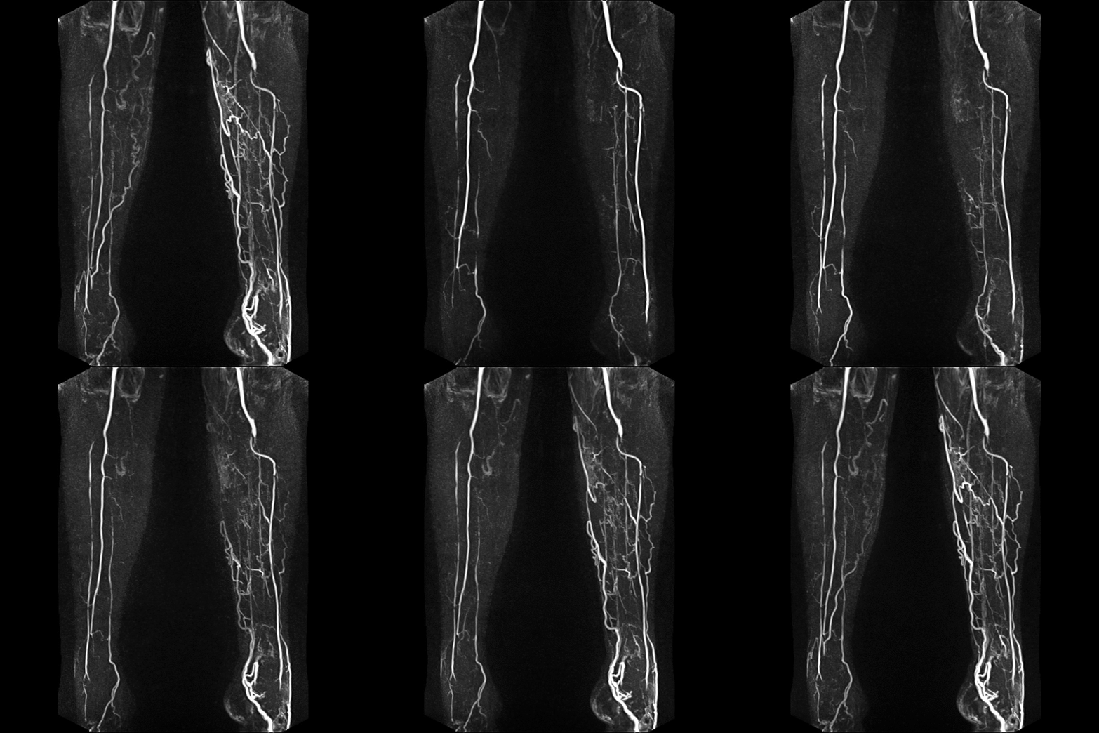
All the contrast-enhanced MRA techniques discussed up to now obtain images at a single point in time after injection. Time-resolved MRA sequences, known under acronyms such as TRICKS and TWIST, obtain a series of images displaying passage of the contrast bolus. A typical time-resolved MRA study might contain 20+ images obtained at rates as rapid as 1-2 frames per second.
An inherent trade-off exists between spatial and temporal resolution. The center of k-space contains information about basic image contrast, while edges and details are encoded in the k-space periphery. Increasing spatial resolution thus requires that more k-space points be sampled. However, sampling more points requires additional imaging time, adversely impacting temporal resolution.
|
|
Time-resolved MRA techniques balance these competing resolution requirements through a process known as view-sharing. Although the details of these methods vary, all begin by acquiring a non-contrast, full-resolution image of the area of interest. During passage of the contrast bolus, the center of k-space is sampled much more frequently than the periphery, which is updated only periodically. The data from the different partial k-space samplings are combined to create a series of time-resolved images with satisfactory spatial resolution. The original non-contrast image can be used as a mask for subtraction to improve vascular conspicuity.
|
Time-resolved CE-MRA methods all trace their origins to a method known as keyhole imaging, described in a prior Q&A. Keyhole imaging was developed in the 1990's and used primarily for performing dynamic contrast-enhanced MR imaging of tumors and vascular lesions. Using a rectangular (Cartesian) k-space grid, the central phase-encoding lines were repeatedly sampled after contrast injection with only occasional sampling of the peripheral lines. View-sharing was used to produce a set of full-resolution images from the data sets.
Modern time-resolved MRA techniques typically use radial sampling schemes, acquiring 3D k-space in round or oval "cylinders". Several vendor-specific implementations are compared below. GE uses the acronym TRICKS ("Time-Resolved Imaging of Contrast KineticS"); Siemens uses TWIST ("Time-resolved angiography With Stochastic Trajectories"), Philips calls theirs 4D-TRAK ("4D Time-Resolved Angiography using Keyhole"), Hitachi uses TRAQ ("Time-Resolved AcQuisition") and Toshiba Freeze Frame.
|
Philips' 4D-TRAK is a centric version of the classic keyhole method, where k-space is divided into a central and peripheral oval regions. Sampling of the central region is acquired much more frequently than the periphery.
Siemens' TWIST also divides k-space into two regions, but samples them alternately using a semi-randomized method. The peripheral region is sparsely sampled at each time point, although is eventually covered over several cycles.
GE's TRICKS divides k-space into four concentric regions (A-D), sampling them in the order
--A-B-A-C-A-D--A-B-A-C-A-D--
|
All time-resolved MRA methods have at their core a 3D-spoiled GRE sequence with thin slices, very short TRs and TEs, low flip angles, use of both read- and phase-conjugate symmetry, parallel imaging acquisition, and zero-interpolation filling in the slice direction. Specific k-space imaging parameters must also be selected. These vary by vendor and may include the size of the central k-space region (typically 15-30%), the undersampling fraction of the outer region (typically 20-30%), central k-space refresh rate (in frames per second), and total number of frames to be acquired (depends on anticipated circulation time).
Although used for dynamic perfusion imaging instead of for MRA, GE's sequence DISCO (DIfferential Subsampling with Cartesian Ordering) shares similar features with TRICKS and TWIST. DISCO subdivides k-space into several annular elliptical regions that are randomly and incompletely sampled together with a central region that is consistently sampled. DISCO is incorporated into a 3D spoiled-GRE core sequence with dual in phase-out of phase echoes allowing separation of fat and water signals. At present, DISCO is primarily used for dynamic contrast-enhanced studies of the breast, liver, and prostate.
|
Time-resolved MRA sequences are widely used wherever circulation is rapid (carotids, cardio-pulmonary system) or unpredictable (extremities). The method is particularly useful for evaluating collateral or retrograde flow around stenoses and in the work up of arteriovenous malformations. Accurate timing of bolus arrival is not required; the technologist simply starts the sequence and runs it until the contrast has passed through the vascular system. Much smaller doses of contrast can also be used than with conventional CE-MRA. Fluoroscopic triggering methods used in conventional CE-MRA are low-resolution, 2D versions of these time-resolved sequences.
|
Advanced Discussion (show/hide)»
No supplementary material yet. Check back soon!
References
Grist TM, Mistretta CA, Strother CM, Turski PA. Time-resolved angiography: past, present, and future. J Magn Reson Imaging 2012; 36:1273-1286. [DOI LINK]
Hennig J, Scheffler K, Laubenberger J, Strecker R. Time-resolved projection angiography after bolus injection of contrast agent. Magn Reson Med 1997; 3:341-345. (Basis of TWIST)
Korosec FR, Frayne R, Grist TM, Mistretta CA. Time-resolved contrast-enhanced 3D MR angiography. Magn Reson Med 1996; 36:345-351. (Description of TRICKS).
Laub G. Kroeker R. syngo TWIST for dynamic time-resolved MR angiography. MAGNETOM Flash 2006; 3;92-95. (Brochure from Siemens explaining TWIST).
Saranathan M, Rettmann DW, Hargreaves BA, et al. DIfferential Subsampling with Cartesian Ordering (DISCO); a high spatio-temporal resolution Dixon imaging sequence for multiphasic contrast enhanced abdominal imaging. J Magn Reson Imaging 2012; 35:1484-92.
van Vaals J, Brummer M, Dixon W, et al. ‘Keyhole’ method for accelerating imaging of contrast agent uptake. J Magn Reson Imaging 1993; 3: 671-5.
Willinek WA, Hadizadeh DR, von Falkenhausen M, et al. 4D time-resolved MR angiography with keyhole (4D-TRAK): More than 60 times accelerated MRA using a combination of CENTRA, keyhole, and SENSE at 3.0T. J Magn Reson Imaging 2008; 27:1455-1460.
Related Questions
How is contrast-enhanced MRA performed?
I know there are different view ordering options for MRA, such as linear and elliptical centric. What do these mean, and when is each used?
How is contrast-enhanced MRA performed?
I know there are different view ordering options for MRA, such as linear and elliptical centric. What do these mean, and when is each used?


