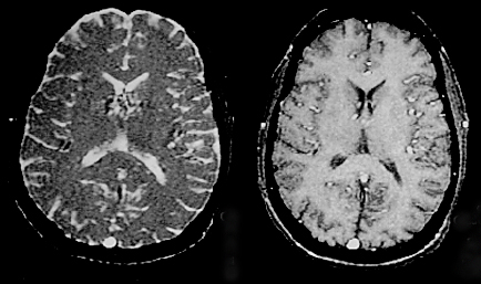
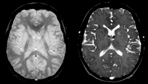
Like most other MRI techniques, image contrast in a GRASS/FISP sequence depends on three operator-selectable parameters (TR, TE, and α) and three tissue-specific parameters (T1, T2*, and [H]. Unlike the spoiled-GRE case, however, the effects of each of these on image contrast is not quite so intuitive nor easily predictable. The signal intensity equation for a GRASS/FISP sequence can be derived by a recursive process with the following formidable result:
The reason this formula is so complex is that both longitudinal and transverse steady-state components are "mixed" or exchanged with each RF-pulse. The final MR signal is therefore a summation of FIDs, secondary spin echoes and stimulated echoes that accumulate over over many cycles. In spite of this complexity, a few basic statements concerning image contrast in these sequences are possible.
TE controls T2*-weighting
Just as with fully spoiled sequences, GRASS/FISP dephases and rephases an FID-like signal into a gradient-recalled echo. This gradient echo exponentially decays with time constant T2*. The longer the echo time (TE), the more time for this decay and lower the overall signal. Just as with spoiled GRE sequences, therefore, lengthening TE in GRASS/FISP increases T2*-weighting.
Long TR or Multi-slice mode: Spoiled-GRE Behavior
|
GRASS/FISP contrast is perhaps the easiest to understand in cases where the assumptions of a coherent transverse steady state break down. For example, when TR is much longer than T2* (e.g., TR > 250 ms for most tissues), natural T2*-processes intrinsically result in complete decay of the transverse magnetization between RF-pulses. The GRASS/FISP sequence then behaves as a spoiled-GRE sequence whose contrast behavior has been described in a separate Q&A.
|
Physiologic motion may also contribute to this intrinsic spoiling of the GRASS/FISP sequence. Thus flowing cerebrospinal fluid and blood may exhibit spoiled contrast behavior, even though stationary tissue remains in a steady-state free precession.
Finally, operating the sequence in multi-slice mode (MPGR) also disrupts the development of coherent transverse magnetization and the sequence develops spoiled-GRE behavior. This will be discussed more fully in the next Q&A.
Finally, operating the sequence in multi-slice mode (MPGR) also disrupts the development of coherent transverse magnetization and the sequence develops spoiled-GRE behavior. This will be discussed more fully in the next Q&A.
Effect of Flip Angle (α)
|
For shorter values of TR, both a longitudinal and a transverse steady state become established with considerable mixing from pulse to pulse. As such, and image contrast is largely independent of TR and will be dictated principally by flip angle (α). As will be explained below, low flip angles (α<20°) produce spin-density-weighted images, whereas larger flip angles (α>45°) result in contrast that is sensitive to the ratio T2/T1.
|
In a prior Q&A I described how a low flip-angle pulse creates an appreciable transverse magnetization while disturbing the longitudinal magnetization only slightly. When α is small, therefore, most of the steady-state magnetization remains in the longitudinal direction, a phenomenon dependent primarily upon the trigonometric fact that cos α ≈ 1 for small α (and hence essentially independent of tissue T1 or T2). Stated another way, the longitudinal magnetization will not change much after a small α pulse, and this is true regardless of what T1 or T2 may be. For low flip angles, therefore, image contrast depends primarily upon the equilibrium magnetization, which reflects the total number of spins in the sample. Hence low flip angles (α < 20°) produce spin-density [H]-weighted images.
When α is large, however, considerable interchange between the longitudinal and transverse magnetization components occurs with each RF-pulse. The net signal then depends strongly upon both T1 and T2 (or more explicitly, upon the ratio T2/T1). For most solid tissues, T1 is approximately 5-10 times longer than T2, but for fluids T2 and T1 are nearly equal. As flip angle increases, therefore, the signal from CSF and other fluids significantly increases because of this T2/T1 behavior.
A summary of these image contrast manipulations appears in the table below. The reader should realize that terms such as "T1-weighting" and "T2*-weighting" are not strictly defined and that in a single image many different tissue contrasts may be present simultaneously. For example, some parts of a GRASS/FISP image may exhibit T1 contrast while other parts show T2/T1 contrast and spin-density weighting. Furthermore, the signal from water/CSF cannot be used as a simple and reliable guide to the type of weighting as it can in SE imaging. Water may be equally bright on spin-density-, T2*-, and T2/T1-weighted steady-state GRE images.
Advanced Discussion (show/hide)»
Additional Notes on GRASS/FISP Contrast
Although the GRASS/FISP signal equation is dominated by its final term (e−T2*/TE), you may have noticed that the factor "A" is a function of T2, not T2*. Thus GRASS/FISP also has some true-T2-weighting in addition to weighting of the T2* variety. The reason for this is that the refocused postexcitation signal sampled in GRASS/FISP is more than a simple FID. In fact, it contains spin echo and stimulated echo contributions from several previous cycles which are affected by true T2 decay.
References
Haacke EM, Frahm J. A guide to understanding key aspects of fast gradient-echo imaging. J Magn Reson Imaging 1991; 1:621-624.
Oppelt A, Graumann R, Barfuss H, et al. FISP: a new fast MRI sequence. Electromedica 1986; 54:15-18.
Haacke EM, Frahm J. A guide to understanding key aspects of fast gradient-echo imaging. J Magn Reson Imaging 1991; 1:621-624.
Oppelt A, Graumann R, Barfuss H, et al. FISP: a new fast MRI sequence. Electromedica 1986; 54:15-18.


