Subclavian Pseudo-stenosis Artifact
|
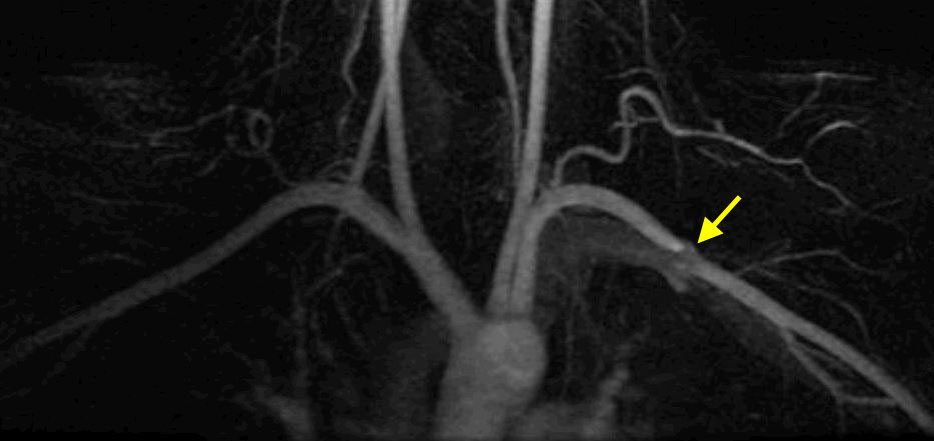
A focal irregularity in the subclavian artery mimicking stenosis may occur ipsilateral to the side of contrast injection. This is a susceptibility (T2*) effect due to residual gadolinium in the adjacent subclavian vein. The artifact is more commonly seen after left-sided injections due to the longer course of the left brachiocephalic vein.
|
|
Scanning Too Late: Venous Contamination
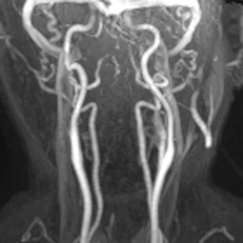
Scanning too late allows time for venous return to occur, overlapping the arterial phase. This is particularly problematic in the intracranial and cervcial carotid systems where significant filling of the dural sinuses and jugular veins may occur within 5-10 seconds after arrival of the contrast bolus. The renal system is another place where rapid venous filling may be witnessed.
Just like CTA, non-time-resolved CE-MRA offers only one chance to "get it right". |
Scanning Too Early: Ringing (Maki) Artifact
|
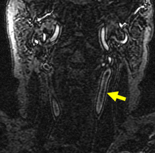
If the central region of k-space is scanned before arrival of the contrast bolus, the vessel will appear dark in the middle, with only its edges demonstrating enhancement. The reason for this appearance follows immediately from the fact that the center of k-space determines basic image contrast while the periphery provides high spatial frequencies and details. This "too-early" phenomenon was originally described by Maki et al. and is called the "Maki" or "ringing" artifact. Like the other artifacts above, the full dose of contrast has generally been used before the artifact is noticed, so no solution is available other than to re-inject the patient, preferably at another sitting.
|
Advanced Discussion (show/hide)»
No supplementary material yet. Check back soon!
References
Lee VS, Martin DJ, Krinsky GA, Rofsky NM. Gadolinium-enhanced MR angiography: artifacts and pitfalls. AJR Am J Roentgenol 2000; 175:197-205.
Maki JH, Prince MR, Londy FJ, Chenevert TL. The effects of time varying intravascular signal intensity and k-space acquisition order on three-dimensional MR angiography image quality. J Magn Reson Imaging 1996;6:642-651.


