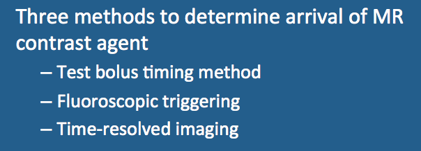
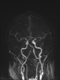
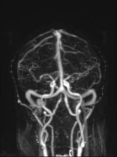
With test bolus timing a small (1-2 mL) test dose of contrast is first administered. The vessel is often imaged in cross section even if a different primary plane of imaging is used for the MRA itself. A series of rapidly acquired 2D images is then obtained over the next 20-40 seconds. These images are examined to determine the time course for passage of the test bolus with calculation of optimal time delay and imaging window. The MRA sequence is then run using these parameters with administration of the remainder (~18 mL) of the gadolinium contrast. The bolus timing method can be performed even on older scanners and provides direct evidence the IV and injector are functioning properly. Disadvantages include 1) increased imaging time; 2) wasting of a small amount of contrast during the test injections; and 3) background contamination by gadolinium (can be severe in renal collecting system and bladder).
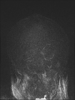
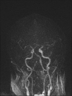
In fluoroscopic triggering the full bolus of contrast (~20 mL) is administered while a fluoroscopic-like picture of the artery of interest is acquired using a rapid 2D gradient-echo technique. Arrival of the bolus is detected by increased signal in the "fluoroscopic" image. At this time the technologist issues a command to begin the MRA imaging acquisition. The fluoroscopic method offers some advantages in that an experienced technologist can watch the passage of contrast through the heart and proximal vessels to judge to optimal time for imaging to begin.
|
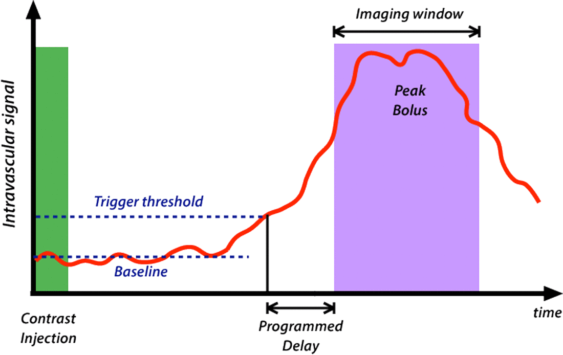
Automatic triggering on bolus arrival can also be performed by placing a region-of interest over the vessel to be imaged. Here the scanner, rather than the technologist, identifies the arrival of the contrast bolus when a specified signal threshold (e.g., 20% above baseline) is exceeded. Once the threshold is reached a programmed delay of 3-4 seconds is interposed to allow the peak bolus to arrive before MRA signal acquisition begins.
|
Advanced Discussion (show/hide)»
The optimal injection rate depends on the clinical application and the type of contrast agent used. As CE-MRA enhancement is due primarily to T1-relaxation effects, agents with higher than average T1-relaxivities (R1), such as gadobuterol (Gadovist) and gadobenate (MultiHance), provide higher levels of arterial signal than the other extracellular agents at standard injection rates of 1.5-3 mL/sec. Gadofosveset (Ablavar), however, because of its albumen binding, has a much more potent T2-relaxivity than other commercially available gadolinium agents. In high concentrations, its T2-shortening competes with T1- shortening and results in reduced intravascular signal. If gadovosveset is used for MRA, therefore, a slower injection rate (1-2 mL/sec) is recommended.
The reader may encounter a number of formulas in the older literature for estimating when to begin data acquisition using the test bolus timing method. One of the most widely used historically was
Td = Tp + ½Ti − ½Ta
where Td = delay time between start of injection and beginning of image acquisition; Tp = time from start of injection to bolus peak, assumed to be equivalent to circulation time; Ti = total injection time, which is assumed to be the same as the time course for contrast filling the artery of interest; and Ta = time for actual MRA data acquisition. Note that this formula applied for scans using linear (sequential) k-space acquisition, where the center of k-space is assumed to be in the center of the data acquisition window. For centric ordering schemes the following equation applies
Td = Tp + Tc
where Tc = time to the center of k-space. Siemens offers a freely adjustable time-to-center parameter on their MRA sequences.
Foo T, Saranathan M, Prince M, Chenevert TL. Automated detection of bolus arrival and initiation of data acquisition in fast, three-dimensional, gadolinium-enhanced MR angiography. Radiology 1997; 203:275-280. (Description of first automated bolus detection method which became GE's SmartPrep)
Menke J. Carotid MR angiography with traditional bolus timing: clinical observations and Fourier-based modelling of contrast kinetics. Eur Radiol 2009; 19:2654-2662.
Wilman AH, Riederer SJ, King BF, et al. Fluoroscopically triggered contrast-enhanced three-dimensional MR angiography with elliptical centric view order: application to the renal arteries. Radiology.1997;205:137–146.
How is contrast-enhanced MRA performed?
I know there are different view ordering options for MRA, such as linear and elliptical centric. What do these mean, and when is each used?
What is bolus-chasing and how is it used for peripheral MR angiography?
