|
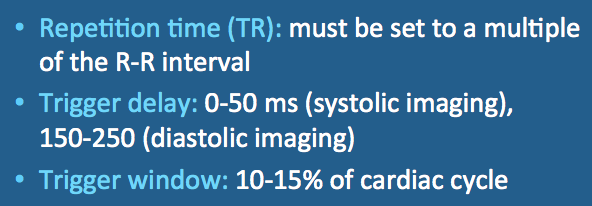
The final 10-15% of each R-R interval is typically reserved as the trigger window. The trigger window serves as a buffer period to allow for slight variations in heart rate. That way, if the next R-wave occurs slightly earlier or later than expected, it will still be detected. R-waves occurring outside the defined trigger window are rejected.
|
Advanced Discussion (show/hide)»
In setting up a cardiac-gated MR scan the technologist reviews the EKG tracings available from various leads and in combination, selecting the one with the most prominent R-wave. Some confusion may arise in that T-waves are often spuriously elevated due to magnetohydrodynamic effects.
Automated detection of the R-wave can be performed based either on greatest amplitude or steepest slope. Several major vendors also offer the option of vector-cardiographic triggering, which is more reliable than simple EKG at distinguishing ventricular ectopic beats and elevated T-waves. The vector-cardiographic method takes into account the angle of the cardiac depolarization vector, which in normal patients is downward and to the left (in the direction of leads I and aVF).
Chia JM, Fischer SE, Wickline SA, Lorenz CH. Performance of QRS detection for cardiac MRI with a novel vectorcardiographic triggering method. J Magn Reson Imaging 2000; 12:678-688.
Comeau CR. Introduction to cardiovascular MR imaging. Milwaukee, GE Medical Systems, 1999. (An old application manual from GE that still contains lots of good basic information).
Lanzer P, Barta C, Botvinick EH, et al. ECG-synchronized cardiac MR imaging: method and evaluation. Radiology 1985;155:681–686.
Niendorf T, Winter L, Frauenrath T. Electrocardiogram in an MRI environment: clinical needs, practical considerations, safety implications, technical solutions, and future directions. In: Mills RM (ed). Advances in electrocardiograms - methods and analysis. InTech Open Access Publishing (www.intech.com), 2012.
Is cardiac gating the same as triggering?


