 Diffusion tensor
Fluids, gels, and homogenous materials can be characterized by a single diffusion coefficient, D, representing the flux of water or small particles via Brownian motion across a surface during a period of time. As described in a previous Q&A, biological tissues are anisotropic with multiple, spatially dependent diffusion coefficients. Diffusion in anisotropic media is better described by the diffusion tensor, a 3x3 matrix containing elements (Dxx, Dxy, Dxz, etc) representing diffusion rates along different directions.
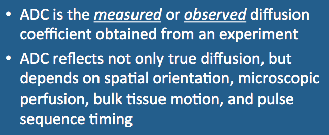
In modern diffusion MR imaging, individual elements of the diffusion tensor are estimated by measuring phase dispersion and signal loss as diffusion-sensitizing gradients are applied in various directions. These methods, however, detect signal loss from several processes in addition to diffusion that can not be easily distinguished — cytoplasmic streaming, blood and lymphatic flow in the microcirculation, bulk tissue motion from cardiac pulsations or respiration, and phase dispersion due to susceptibility effects. Signal losses and diffusion tensor estimates also vary according to the details of the pulse sequence and timing parameters employed.
Chemists in the early 1930's recognized that the measured diffusion rates of dyes and other small molecules were highly dependent both on the nature of the media studied and on experimental conditions. Recognizing these limitations, they began to use the term apparent diffusion coefficient (ADC) to refer to estimated diffusion rates obtained from their studies.
Chemists in the early 1930's recognized that the measured diffusion rates of dyes and other small molecules were highly dependent both on the nature of the media studied and on experimental conditions. Recognizing these limitations, they began to use the term apparent diffusion coefficient (ADC) to refer to estimated diffusion rates obtained from their studies.
|
In the modern MR literature, the term "ADC" is used in at least two different contexts. In the most general sense "ADC" is synonymous with "measured/observed" diffusion rates or tensor components, reflecting the methodological uncertainties described above. In other contexts, "ADC" is used to refer to the mean diffusion in a voxel, sometimes taken as the sum or average value of the tensor's diagonal elements. The commonly viewed ADC map reflects this latter definition.
|
Advanced Discussion (show/hide)»
No supplementary material yet. Check back soon!
References
Le Bihan D. The 'wet mind': water and functional neuroimaging. Phys Med Biol 2007;52:R57-R90.
Le Bihan D, Breton E, Lallemand D, et al. MR imaging of intravoxel incoherent motions: applications to diffusion and perfusion in neurologic disorders. Radiology 1986; 161:401-407.
Le Bihan D, Breton E, Lallemand D, et al. Separation of diffusion and perfusion in intravoxel incoherent motion MR imaging. Radiology 1988;168:497-505.
Minati L, Weglarz WP. Physical foundations, models, and methods of diffusion magnetic resonance imaging of the brain: a review. Concepts Magn Reson 2007; 30A:278-307.
Related Questions
What is diffusion?
How do you make a DW image?
What is the Trace DW image? How does it differ from the ADC map?
What is diffusion?
How do you make a DW image?
What is the Trace DW image? How does it differ from the ADC map?

