|
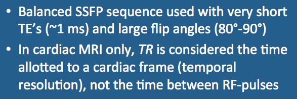
The use of balanced (symmetric) imaging gradients allows simultaneous refocusing of spin echoes, gradient echoes, and stimulated echoes exactly at the midpoint between RF-pulses. The echo time (TE) is made as short as possible, which on modern systems can be in the 1-2 ms range. RF-flip angle (α) is made large (80°-90°) to accentuate T2/T1 contrast and produce bright blood images.
|
|
The TR value used in cardiac MRI is the time allotted to recording data from an each cardiac frame (segment). If, for example, 7 lines of k-space are acquired for each cardiac frame, the "reported" TR would be 7x the "true" TR. Although called both are called "repetition times", I prefer to think of the "reported" TR used in cardiac MRI as an abbreviation for "temporal resolution."
|
|
A trade-off exists between temporal and spatial resolution. The number of cardiac frames (segments) acquired during a single R-R interval and the number of lines per segment are interrelated. As can be seen in the diagram left, if more cardiac phases are desired, fewer lines of k-space can be sampled within each frame, resulting in increased acquisition time.
|
|
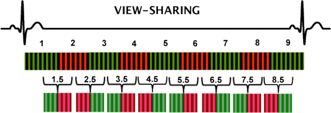
More cardiac phases can be generated by reusing data from adjacent frames through a process known as view-sharing. As seen in the diagram left, interpolated frames labelled 1.5, 2.5, 3.5, etc. can be created by combining data from neighboring segments.
|
Advanced Discussion (show/hide)»
For cine cardiac imaging at 1.5T we currently currently use the following parameters: echo time (TE) = 1.15 ms, flip angle (α) = 90°, slice thickness = 7 mm, number of excitations (NEX) = 1, imaging matrix 156 x 192, and parallel imaging acceleration factor (R) = 2. Depending on heart rate the temporal resolution or "reported" TR is about 38.5 ms. The number of segments is 14, with view sharing allowing reconstruction to 26 cardiac phases for displaying the cine loop.Total imaging time is less than 6 seconds for each anatomic location chosen.
Although FLASH imaging has largely been abandoned in favor of b-SSFP sequences for cardiac cine, occasionally they are still useful. This occurs in the setting of metal valves and stents where susceptibility artifacts may result in poor SSFP images with zebra-stripe phase artifact. For these situations we use a retrospectively gated segmented FLASH cine sequence with the following parameters: "True" TR = 7-10 ms, TE = 2-3 ms, α = 15-20º, views per frame = 3-7, imaging matrix = 128 x 256, NEX = 1. Total acquisition time depending on heart rate is in the 15-20 sec range.
Atkinson DJ, Edelman RR. Cineangiography of the heart in a single breath hold with a segmented TurboFLASH sequence. Radiology 1991; 178:357-360. (In the 1990s and early 2000s, cine studies were performed using a spoiled GRE sequence as in the method described here).
Bieri O, Scheffler K. Fundamentals of balanced steady state free precession MRI. J Magn Reson Imaging 2013;38:2-11.
Carr JC, Simonetti O, Bundy J, et al. Cine MR angiography of the heart with segmented true fast imaging with steady-state precession. Radiology 2001; 219:828-834. (First report of a TrueFISP cine sequence, showing its superiority over segmented TurboFLASH methods).
What is True FISP, and why is it "truer" than regular FISP?
How do they make those movies of the beating heart?


