|
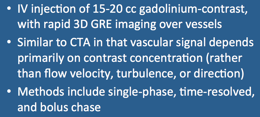
Contrast-enhanced MR angiography (CE-MRA) is similar to contrast-enhanced CT angiography, except a gadolinium-based agent (instead of an iodine compound) is injected. Just as iodine produces x-ray attenuation allowing visualization of vessels on CTA, gadolinium shortens the T1 of blood rendering vessels bright on CE-MRA. Flow-related phenomena (e.g., velocities, flow direction, turbulence) — factors that play dominant roles in the appearance of vessels on non-contrast MRA studies — have little or no effect on CE-MRA where vascular signal intensity is determined principally the the concentration of gadolinium within the vessel.
|
|
The gadolinium-based contrast agent is injected intravenously, usually into a central catheter or antecubital vein. In an average-sized adult with normal renal function, about 20 mL of contrast is administered over 8-12 sec, followed by a saline flush ("chaser") of another 20-30 mL. The goal is to inject a compact bolus of contrast into the central circulation with clearing of all contrast from the proximal veins.
The injection of contrast (and saline) can be performed by hand. This can be done using two syringes and a stop-cock, or with a single syringe loaded with (denser) gadolinium on the bottom and (lighter) saline on top. More commonly power injectors using two syringes remotely controlled from the scanner console are employed.
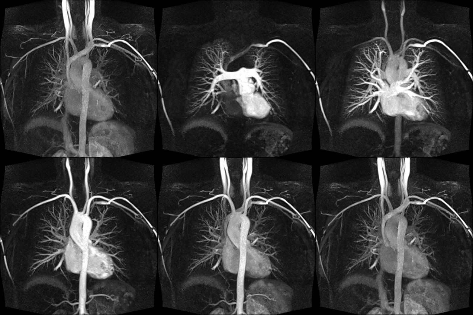
During its passage through the heart and lungs, the contrast agent becomes progressively diluted. For example, if the gadolinium contrast injected into the arm has a concentration of 500 mM, by the time it has reached the carotid arteries its concentration has fallen to less than 10 mM.
|
|
Once the contrast bolus arrives in the vessel of interest, imaging is typically performed using a rapid 3D T1-weighted spoiled gradient-echo pulse sequence. TR and TE are generally made as short as possible, and gradient-moment nulling is not used. A wide receiver bandwidth (equivalent to short sampling time) is typically employed in conjunction with partial k-space acquisition and parallel imaging.
Two different acquisition modes are available. Single-phase methods capture a vascular images at a single point in time. Time-resolved methods repeatedly acquire views of the imaging volume as contrast passes through the regional circulation. Bolus-chase methods combine table movement with imaging to follow the contrast distally into the extremities. |
Advanced Discussion (show/hide)»
If possible right arm veins are preferred for contrast injection as they offer a straighter path to the right heart allowing for a more coherent bolus. Additionally residual contrast in the right innominate vein is less likely to interfere with visualization of aortic arch vessels than contrast remaining in the longer left brachiocephalic system.
Vascular signal intensity in a CE-MRA sequence depends on the arterial gadolinium concentration [Gd]a at each point in time. [Gd]a, depends on the size and shape of the contrast bolus, which is, in turn, related directly to the contrast infusion rate (IR) and inversely to the cardiac output (CO).
[Gd]a ≈ IR/CO
This equation makes since and has practical application. A faster injection rate (IR) delivers a higher and tighter peak bolus of gadolinium and causes more enhancement. Patients with lower cardiac ouputs (CO) will have less dispersion of the contrast bolus and hence greater degrees of arterial enhancement for a given injection rate.
Maki JH, Knopp MV, Prince M. Contrast-enhanced MR angiography. Appl Radiol 2003 (April):182-210.
Medrad Spectris Solaris EP Power Injector (pdf). Product brochure from Bayer HealthCare (www.bayerhealthcare.com), December, 2012.
Prince MR. Gadolinium-enhanced MR aortography. Radiology 1994; 191:155-164. [DOI Link]
Riederer SJ, Haider CR, Borisch EA, et al. Recent advances in 3D time-resolved contrast-enhanced MR angiography. J Magn Reson Imaging 2015; 42:3-22. (good recent review) [DOI Link]
Riederer SJ, Stinson EG, Weavers PT. Technical aspects of contrast-enhanced MR angiography: current status and new applications. Magn Reson Med Sci 2018; 17:3-12. (includes description of newer Dixon CE-MRA and compressed sensing methods) [DOI LINK]
What artifacts on contrast-MRA should we know about?
How do you compute the arrival of contrast in a vessel to know when to start the MR acquisition?
I've heard about using the iron supplement ferumoxytol as an MR contrast agent. What is it and how does it work?


