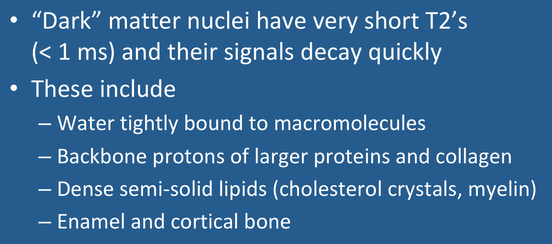
As described in the prior Q&A, nearly all the signals we record from solid organs using conventional MRI originate either from water or triglyceride protons. These ¹H nuclei are relatively abundant and possess T2 relaxation times ranging from tens to hundreds of milliseconds.
 Ring of dark matter in
galaxy cluster CL0024+17
But there is another pool of ¹H nuclei, including water tightly bound to macromolecules, as well as protons along the backbone of larger proteins, solid fats, urate and hydroxyapatite crystals, enamel, cortical bone, collagen, and myelin, whose motions are highly restricted resulting in T2-values measured in microseconds. Their signals decay so quickly that they are not recorded using standard MR techniques and can be considered "MR invisible". I commonly refer to them as "dark protons" or the "dark pool". Our current failure to routinely visualize these dark protons implies we are missing out on a significant amount of potentially important information!
Like dark matter in the universe, dark protons are ubiquitous and may constitute at least 20% of ¹H nuclei potentially available for MR analysis in most solid organs. Those with super-short T2's (<0.1 ms) are currently not detectable with any clinical sequences, but can be measured indirectly through their interaction with other visible protons though dipolar and magnetization transfer mechanisms. Protons with slightly longer T2's (0.1 - 1.0 ms) can be rendered visible with a new class of ultrashort TE sequences (including UTE, PETRA, and ZTE), the subject of several later Q&As. Myelin (whose average T2 is about 0.3 ms) is an important component of this second category and which can now be visualized imaged using specialized UTE sequences (e.g. DESIRE, STAIR) that suppress unwanted signals from longer T2 components.
An intermediately dark pool of protons, up to 40% by weight, are present in cartilaginous/fibrous tissues, such as ligaments, menisci, tendons, and periosteum. Their average T2 values lie in the 1-10 ms range, making them relatively "invisible" on long TE sequences, but detectable by short TE sequences achievable by most current clinical scanners. Because these tissues contain organized strands of collagen and/or elastin, both relaxation times and susceptibilities vary according to orientation in the main magnetic field, the so-called magic angle phenomenon which must be taken into account when imaging them.
Advanced Discussion (show/hide)»
No supplementary material yet. Check back soon!
References
Bydder GM. The Agfa Mayneord lecture: MRI of short and ultrashort T2 and T2* components of tissues, fluids, and materials using clinical systems. Brit J Radiol 2011; 84:1067-1082. (The first published reference I can find where these protons are referred to as "dark matter").
Chang EY, Du J, Chung CB. UTE imaging in the musculoskeletal system. J Magn Reson Imaging 2015; 41:870-883.
Halle B. Water in biological systems: the NMR picture. In: Bellissent-Funel M-C (ed.). Hydration processes in biology : theoretical and experimental approaches. IOS Press, Clifton, VA. 1999: 233-249.
Horch RA, Nyman JS, Gochberg DF, et al. Characterization of ¹H NMR signal in human cortical bone for magnetic resonance imaging. Magn Reson Med 2010; 64:680-7.
Wilhelm MJ, Ong HH, Wehrli SL, et al. Direct magnetic resonance detection of myelin and prospects for quantitative imaging of myelin density. Proc Natl Acad Sci (USA) 2012; 109:9605-9610.
Related Questions
I'm getting confused. You've been talking about all kinds of nuclei -- in fat, water, and macromolecules. Which hydrogen protons are producing the MR signal?
What is the magic angle?
I'm getting confused. You've been talking about all kinds of nuclei -- in fat, water, and macromolecules. Which hydrogen protons are producing the MR signal?
What is the magic angle?
