|
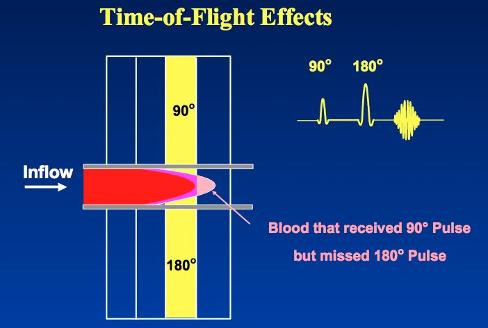
Time-of-flight (TOF) effects refer to signal variations resulting from the motion of protons flowing into or out of an imaging volume during a given pulse sequence. In both spin-echo and gradient-echo imaging, inflow of spins results in increased signal; this phenomenon is known as flow-related enhancement. Conversely, outflow of spins may result in decreased signal intensity, a phenomenon known as high-velocity signal loss, also called "washout".
|
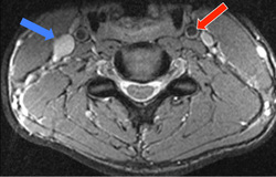
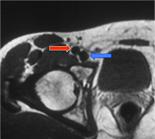
Both TOF effects are demonstrated in the image above. The more slowly flowing blood in the jugular vein (blue arrow) demonstrates flow-related enhancement. The more rapidly flowing blood in the carotid artery (red arrow) has low signal due to flow-related signal loss (washout).
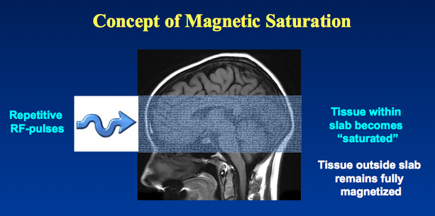
Flow-Related Enhancement. Flow-related enhancement is a consequence of magnetic saturation, a phenomenon described more completely in a prior Q&A and illustrated in the diagram below. Tissues contained within an imaging volume are subjected to a continuous train of radiofrequency (RF) pulses. These RF pulses repeatedly flip the longitudinal tissue magnetization (Mz) into the transverse plane. If the pulses are widely spaced (TR > 5 x T1), complete recovery occurs and no saturation effect is observed.
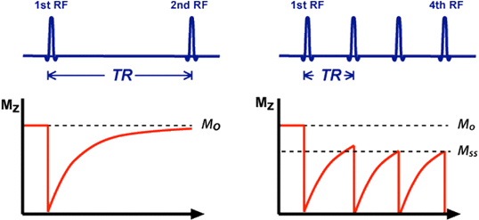
Partial saturation and development of steady-state magnetization (Mss)
[left] TR >> T1 so there is complete recovery of Mz back to Mo before the 2nd RF-pulse.
[right] TR is shorter and Mz does not fully recover before next RF-pulse is applied. After a few pulses a new steady-state longitudinal magnetization (Mss) is reached, with Mss < Mo.
[left] TR >> T1 so there is complete recovery of Mz back to Mo before the 2nd RF-pulse.
[right] TR is shorter and Mz does not fully recover before next RF-pulse is applied. After a few pulses a new steady-state longitudinal magnetization (Mss) is reached, with Mss < Mo.
Most sequences, however, utilize shorter TR values, and hence the next RF-pulse is applied before Mz has fully recovered. The longitudinal magnetization evolves to a new lower steady state (Mss). The tissues within the imaged volume are said to be partially saturated, as their MR signals are lower than they would otherwise be.
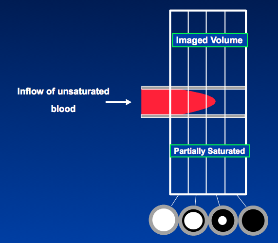
"Fresh" blood flowing into the imaged slab has not been subjected to these RF-pulses and is therefore fully magnetized (unsaturated). When a bolus of this fresh blood enters a slice and is subjected to its first set of RF-pulses, its signal is significantly stronger than that of the tissues (and other blood) within the slab. This is the genesis of flow-related enhancement.
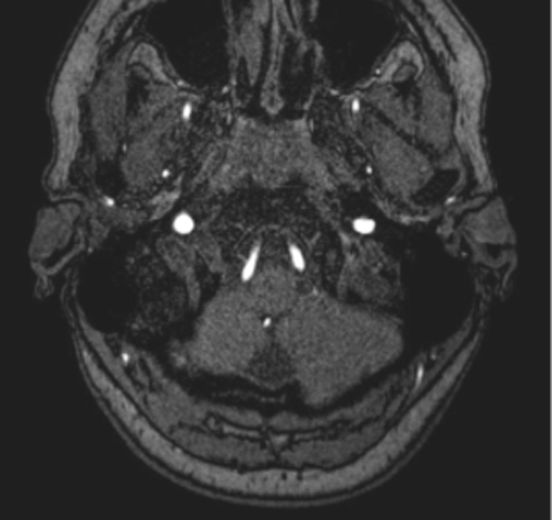
Flow-related enhancement is typically seen at the end slices of a multi-slice acquisition where the fresh blood is entering. If blood flow is sufficiently rapid, this effect may penetrate several slices deep into the slab. Sometimes only a central bright dot will be seen in the vessel at the deeper slices corresponding to the fastest flow in the center of the vessel.
|
High-velocity signal loss. High-velocity signal loss, also known as "washout", is a time-of-flight effect that results in decreased intravascular signal. This phenomenon occurs especially in spin-echo imaging and is typically seen in arteries and larger veins that are flowing perpendicular plane of imaging. Unlike flow-related enhancement, high-velocity signal loss can occur anywhere in the imaging slab and is not confined to the end slices.
|
High-velocity signal loss occurs when spins flow out of slice before they are stimulated by both 90° and 180° pulses. It is therefore most prominent on long TE spin-echo images, since long echo times provide a greater opportunity for spins to flow out of the slice between the 90° and 180° pulses.
|
Advanced Discussion (show/hide)»
Historically, flow-related enhancement was sometimes called "paradoxical" enhancement, but this term has largely fallen into disuse.
The reason high-velocity signal loss does not occur on GRE imaging is that the refocusing mechanism (gradient reversal) is not slice selective.
References
Axel L. Blood flow effects in magnetic resonance imaging. AJR Am J Roentgenol 1984; 143:1157-1166.
Bradley WG Jr, Waluch V. Blood flow: magnetic resonance imaging. Radiology 1985; 154:443-450. Bradley WG Jr, Waluch V, Lai K-S, et al. The appearance of rapidly flowing blood on magnetic resonance images. AJR Am J Roentgenol 1084; 143:1167-1174.
Wehrli FW. Time-of-flight effects in MR imaging of flow. Magn Reson Med 1990; 14:187-193.
Related Questions
Wait a minute! If a partial flip angle pulse tips less magnetization into the transverse plane than a 90° pulse does, how can the signal be higher?
Wait a minute! If a partial flip angle pulse tips less magnetization into the transverse plane than a 90° pulse does, how can the signal be higher?

