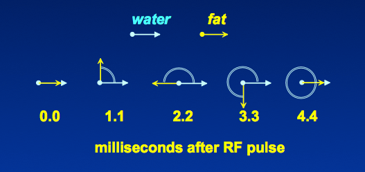
As described in the prior Q&A, phase cancellation between water and fat signals arising from the same voxel gives rise to a specific artifact known as the chemical shift artifact of the second kind on gradient echo images. This artifact is manifest by a black line around organs and anatomic structures at water-fat interfaces. The magnitude of this artifact depends on the phase cycling between water and fat, which because of their chemical shifts, resonate with a frequencies varying by about 215 Hz at 1.5T.
|
This phase cycling between in-phase and out-of-phase occurs approximately every 2.2 msec at 1.5T. (At 3.0T the cycling is twice as fast, occurring every 1.1 msec). GRE images obtained at 1.5T at TE's of 2.2, 6.6, 11.0 msec are called out-of-phase (OOP); those obtained at 4.4, 8.8, etc. are called in-phase (IP).
|
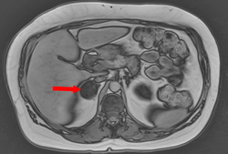
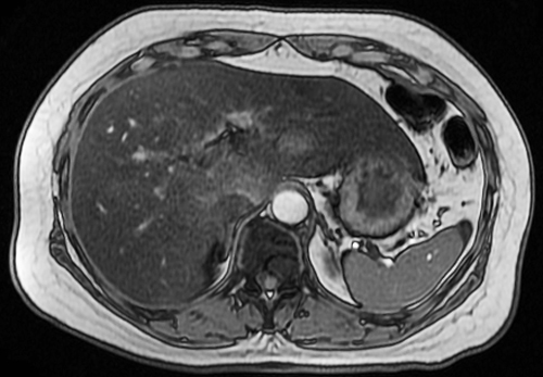
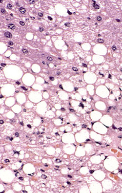
By the late 1980's several investigators began to realize that this phase cancellation effect could be used clinically to identify and even quantify the fat content of tissues like the liver. One particularly common use of this principle today is to help in the differentiation of adrenal adenomas (that typically contain fat) from carcinomas and metastases (that do not). The diagnosis of a variety of other abdominal lesions, including angiomyolipomas, renal clear cell carcinoma, and focal fatty infiltration of the liver may be assisted by IP-OOP imaging. The technique, illustrated below, involves obtaining a pair of GRE images at the same TR but with two different TE values, one IP and the second OOP. Lesions whose signal intensities drop significantly on the OOP images are likely to contain microscopic fat. Accordingly, IP/OOP scanning is now a standard part of most abdominal imaging protocols world-wide.
|
|
|
Advanced Discussion (show/hide)»
No supplementary material yet. Check back soon!
References
Outwater EK, Blasbalg R, Siegelman ES, Vala M. Detection of lipid in abdominal tissues with opposed-phase gradient-echo images at 1.5T: techniques and diagnostic importance. Radiographics 1998; 18:1465-80.
Outwater EK, Blasbalg R, Siegelman ES, Vala M. Detection of lipid in abdominal tissues with opposed-phase gradient-echo images at 1.5T: techniques and diagnostic importance. Radiographics 1998; 18:1465-80.
Related Questions
What is a chemical shift artifact of the second kind?
How do you produce multiple GRE's from a single pulse?
What is a chemical shift artifact of the second kind?
How do you produce multiple GRE's from a single pulse?


