Note: In this Q&A we adopt the convention that substances "bright" on T1-weighed images have short T1 values. This is true for most (but not all) pulse sequences. For a detailed explanation, click here.
|
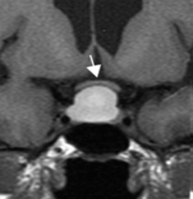
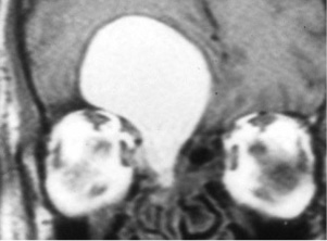
Several clinical entities are commonly recognized where T1 shortening (and high T1 signal) occurs due to high concentrations of macromolecules. Examples include mucoceles, colloid cysts, Rathke cleft cysts, neuroenteric cysts, bronchogenic cysts, renal cysts, biliary sludge, pigment stones, and "white" epidermoids.
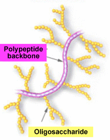
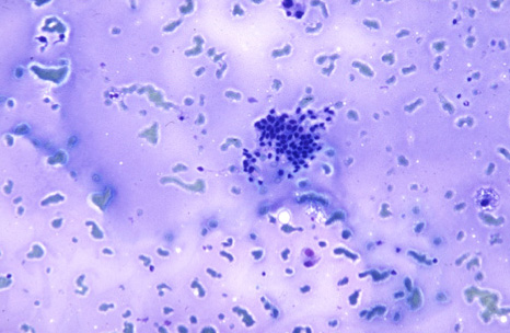
Mucin. By weight composition normal mucus of the aerodigestive tract is approximately 95-98% water and 2-5% solids, with about 60% of the solid, high molecular weight component (mucin) formed by a polypeptide backbone with attached oligosaccharides. These side chains are responsible for the short T1 values either by themselves or from water-macromolecule interactions.
As an interesting aside, many of these sugars are N-acetyl linked (GalNAc, GlcNAc). Hydrogen MRS of mucoceles may reveal a distinct peak at δ = 2.0, mimicking the NAA peak seen in normal brain. |
|
Due to its high water content, mucus usually has CT and MR characteristics similar to water. Mucinous cystadenomas and cystadenocarcinomas, for example, even those with >50% mucin content, generally are not bright on T1-weighted images. However, concentrated mucus may have high attenuation values on CT (up to 130 HU) and short T1 relaxation times. A protein level of > 9 g/dl is generally required to produce high signal on T1-weighted images, but much lower concentrations give mucin-containing structures a high enough T1 signal to distinguish them from pure water. The relaxation characteristics of intraluminal mucin also depends on whether it is in more of a liquid or gelatinous/viscous phase as well as whether coexisting debris or cellular material is also present.
|
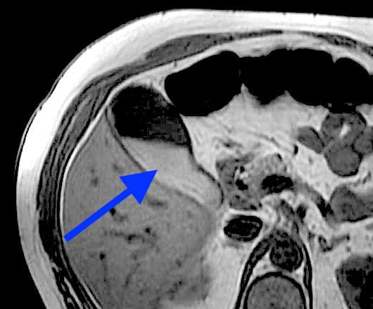
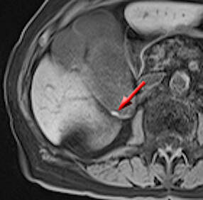
Hyperintense signal on T1-weighted images is commonly seen in concentrated bile and gallbladder sludge. Although biliary cholesterol has been cited as the reason for T1-shortening, this has been disproved by several ¹H MR spectroscopy studies showing that nearly all of the MR signal in bile comes from water, not lipid protons. Mucin, however, is found in high concentrations in biliary sludge, and a water-mucin interaction seems the most likely cause. Likewise, pigmented (but not cholesterol) gallstones are bright on T1-weighted MR images, probably because they are formed on a mucin-glycoprotein matrix constituting about 25% of the stone by weight.
|
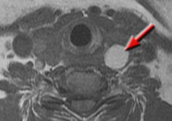
Colloid. The term colloid is non-specific and refers to a wide range of emulsions, gels, and viscous liquids whose chemical content may differ. In the thyroid, for example, colloid is generally synonymous with concentrated thyroglobulin, a 660 kDa glycoprotein containing T3 and T4 hormones. The relaxation time of various colloids will thus vary by concentration and type of substances contained therein.
|
|
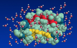
The physics behind T1 shortening of solutions high in proteins and/or macromolecules is more completely described in a separate Q&A (click here for link). In brief, bonding between water and macromolecular hydrogens occurs at the interface, slowing correlation times so that a higher fraction of water protons rotate near the Larmor frequency (making T1 relaxation more efficient). Additionally, chemical exchange results in magnetization transfer between the two proton pools.
|
Advanced Discussion (show/hide)»
No supplementary material yet. Check back soon!
References
Gaeta M, Vinci S, Minutoli F, et al. CT and MRI finding of mucin-containing tumors and pseudotumors of the thorax: pictorial review. Eur Radiol 2002; 12:181-189.
LaMont JT, Ventola AS, Trotman BW, Soloway RD. Mucin glycoprotein content of human pigment gallstones. Hepatology 1983; 3:377-382.
Lee NK, Kim S, Kim HS, et al. Spectrum of mucin-producing neoplastic conditions of the abdomen and pelvis: cross-sectional imaging evaluation. World J Gastroenterol 2011; 17:4757-4771
Ochi M, Hayashi K, Hayashi T, et al. Unusual CT and MR appearance of an epidermoid tumor of the cerebellopontine angle. AJNR Am J Neuroradiol 1988; 19:1113-5.
Ostrow JD. The etiology of pigment gallstones. Hepatology 1984; 4:215S-222S.
Tsai H-M, Lin X-Z, Chen C-Y et al. MRI of gallstones with different compositions. AJR 2004; 182:1513-1519.
Related Questions
What is T1 relaxation?
Can you explain a little more about the dipole-dipole interaction? I still don't quite understand.
What is T1 relaxation?
Can you explain a little more about the dipole-dipole interaction? I still don't quite understand.

